Copyright
©The Author(s) 2015.
World J Nephrol. Sep 6, 2015; 4(4): 455-467
Published online Sep 6, 2015. doi: 10.5527/wjn.v4.i4.455
Published online Sep 6, 2015. doi: 10.5527/wjn.v4.i4.455
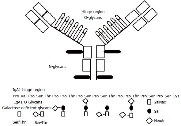
Figure 1 Immunoglobulin A1 and its hinge region with O-linked glycans (white) and N-linked glycans (black).
Under the aminoacids chain of the hinge region. Sites of attachment are in bold. IgA: Immunoglobulin A.
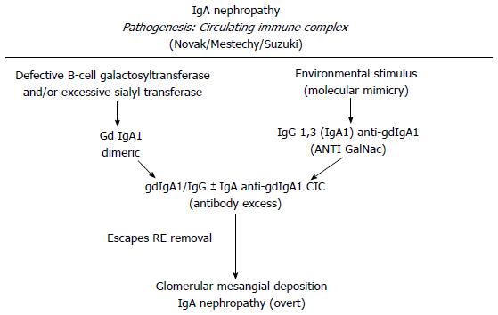
Figure 2 Schematic representation for the possible pathways involved in the generation of a circulating immune complex pathogenesis for the immunoglobulin A nephropathy.
CIC: Circulating immuno complex; Gd IgA1: Galactose deficient immunoglobulin A1.
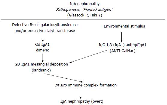
Figure 3 Schematic representation for the possible pathways involved in an in situ pathogenesis for the IgA nephropathy.
IgA: Immunoglobulin A; Gd-IgA1: Galactose deficient immunoglobulin A1; ANTI GalNac: ANTI N-acetylgalactosamine.
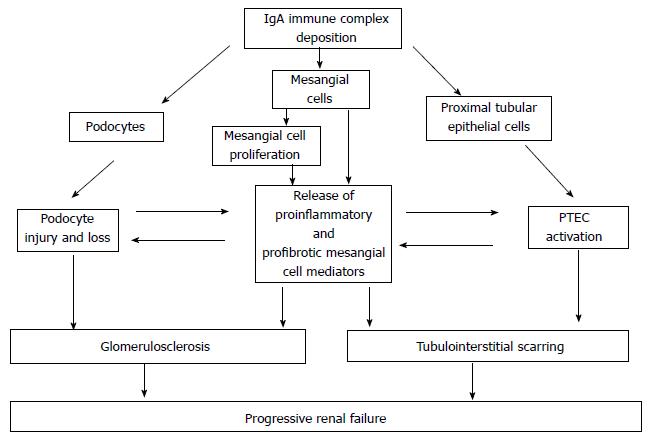
Figure 4 Pathways to glomerular damage and tubulointerstitial injury in Immunoglobulin A nephropathy.
Deposition of IgA-ICs in the mesangium leads to activation of mesangial cells, triggering mesangial cell proliferation and release of proinflammatory and profibrotic mediators. Podocyte loss accentuates glomerular scarring and filtered mesangial cell-derived mediators and IgA-ICs stimulate PTEC to adopt a proinflammatory and profibrotic phenotype, which in turn drives tubulointerstitial scarring. IgA: Immunoglobulin A; PTEC: Proximal tubule epithelial cells.
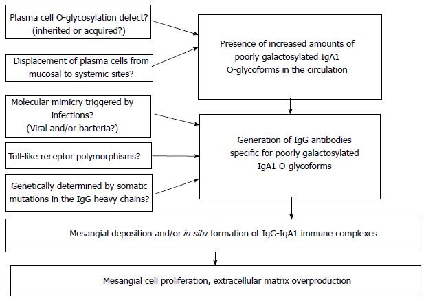
Figure 5 Doubts and different possibilities in generating the first two steps.
IgA: Immunoglobulin A; IgG-IgA1: Immunoglobulin G-Immunoglobulin A1.
- Citation: Salvadori M, Rosso G. Update on immunoglobulin A nephropathy, Part I: Pathophysiology. World J Nephrol 2015; 4(4): 455-467
- URL: https://www.wjgnet.com/2220-6124/full/v4/i4/455.htm
- DOI: https://dx.doi.org/10.5527/wjn.v4.i4.455


