Copyright
©The Author(s) 2015.
World J Nephrol. May 6, 2015; 4(2): 313-318
Published online May 6, 2015. doi: 10.5527/wjn.v4.i2.313
Published online May 6, 2015. doi: 10.5527/wjn.v4.i2.313
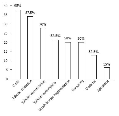
Figure 1 Percentage of the elementary lesions in the kidney.
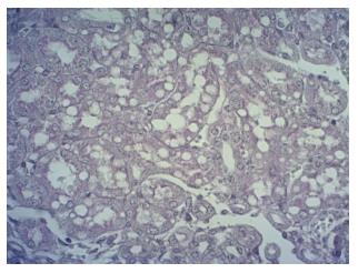
Figure 2 Vacuolation of the proximal tubules.
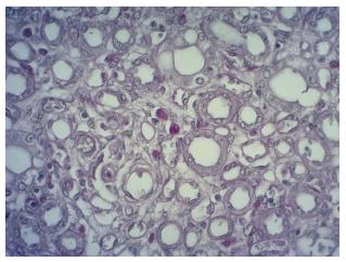
Figure 3 Apoptosis.
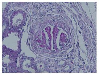
Figure 4 Fibrous and cellular crescents associated with collapse of the glomerular tuft.
- Citation: Gerosa C, Iacovidou N, Argyri I, Fanni D, Papalois A, Aroni F, Faa G, Xanthos T, Fanos V. Histopathology of renal asphyxia in newborn piglets: Individual susceptibility to tubular changes. World J Nephrol 2015; 4(2): 313-318
- URL: https://www.wjgnet.com/2220-6124/full/v4/i2/313.htm
- DOI: https://dx.doi.org/10.5527/wjn.v4.i2.313


