Copyright
©2014 Baishideng Publishing Group Inc.
World J Nephrol. Nov 6, 2014; 3(4): 302-307
Published online Nov 6, 2014. doi: 10.5527/wjn.v3.i4.302
Published online Nov 6, 2014. doi: 10.5527/wjn.v3.i4.302
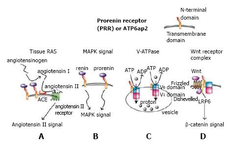
Figure 1 Four roles of prorenin receptor.
A: When renin or prorenin binds to prorenin receptor (PRR), renin or prorenin enzymatic activity is enhanced through non-proteolytic conformational change, catalyzing angiotensinogen to angiotensin I. Produced angiotensin I is catalyzed by angiotensin-converting enzyme, yielding angiotensin II that induces angiotensin II receptor-mediated signal transduction, ending in enhanced tissue renin-angiotensin system (RAS)[45]; B: When PRR is bound to a ligand, renin, or prorenin, a mitogen-activated protein kinase (MAPK) signal is induced[1]; C: PRR, with or without the N-terminal domain, is required as a subunit of V-ATPase, which actively transports protons into vesicles such as endosomes, lysosomes, and autophagosomes using energy obtained by degrading ATP to ADP[9]. The V0 and V1 domains build up V-ATPase; D: PRR is required as an adaptor protein between V-ATPase and LRP6, which are members of the Wnt receptor complex[5,46].
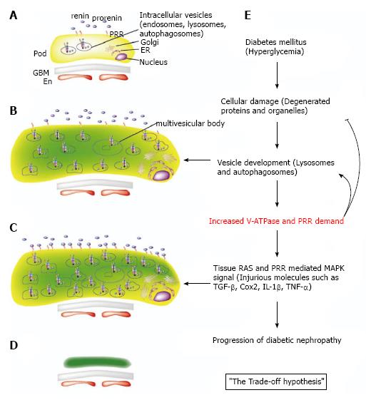
Figure 2 The trade-off hypothesis in diabetic nephropathy from the prorenin receptor perspective.
A: Normal podocyte intracellular structure; B: Under hyperglycemic conditions, injured podocytes degrade degenerated proteins and intracellular organelles, forming vesicles such as lysosomes and autophagosomes, which require V-ATPase and prorenin receptor (PRR) for internal acidification; C: Sustained hyperglycemia overproduces PRR molecules, which are transported to the transmembrane and bind to increased serum prorenin in the diabetic condition. This enhances tissue renin-angiotensin system (RAS) and PRR-mediated mitogen-activated protein kinase (MAPK) signals; D: Atrophic or apoptotic podocytes after long-term hyperglycemic conditions. The changes in the glomerular basement membrane and endothelial cells are not shown; E: The schematic view of the trade-off hypothesis. In diabetes mellitus, podocyte injury occurs by producing degenerated proteins and organelles, which in turn are degraded in lysosomes and autophagosomes. This process requires V-ATPase and PRR production for internal acidification of the vesicles. PRR overproduction enhances tissue RAS and PRR-mediated MAPK signals, resulting in increased injurious molecules such as transforming growth factor-β, cyclooxygenase2, interleukin-1β, and tumor necrosis factor-α. Progression and deterioration of diabetic nephropathy occurs at the end. En: Endothelial cell; ER: Endoplasmic reticulum; GBM: Glomerular basement membrane; Pod: Podocyte.
- Citation: Oshima Y, Morimoto S, Ichihara A. Roles of the (pro)renin receptor in the kidney. World J Nephrol 2014; 3(4): 302-307
- URL: https://www.wjgnet.com/2220-6124/full/v3/i4/302.htm
- DOI: https://dx.doi.org/10.5527/wjn.v3.i4.302


