Copyright
©2013 Baishideng Publishing Group Co.
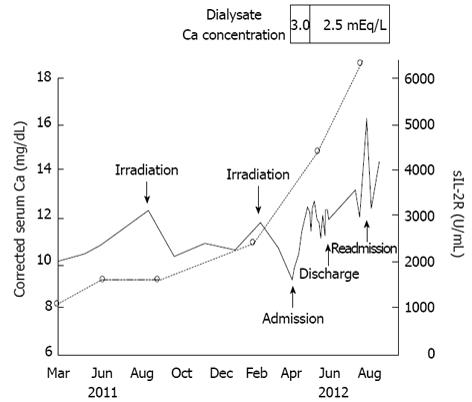
Figure 1 Clinical course.
The solid line indicates changes in corrected serum calcium concentrations. The dotted line indicates changes in soluble interleukin-2 receptor levels. The corrected serum calcium concentration was calculated as follows: corrected serum calcium concentration (mg/dL) = serum calcium concentration (mg/dL) + [4-serum albumin concentration (mg/dL)]. sIL-2R: Soluble interleukin-2 receptor.
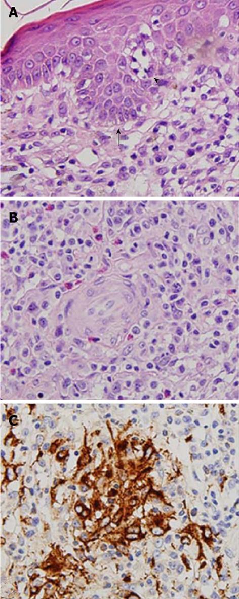
Figure 2 Microphotographs of skin biopsy specimens.
A: There are clusters of atypical lymphocytes that form a Pautrier microabscess (arrow) within the epidermis and diffuse and dense small to medium-sized atypical lymphocytes with hyperchromatic and convoluted nuclei throughout the whole dermis. Epidermotropism of lymphocytes (arrow head) is seen. (Original magnification: × 400. Periodic acid-Schiff staining); B: Granuloma formation with aggregation of histiocytes is seen, and multinucleated histiocytic giant cells are present. (Original magnification: × 400. Periodic acid-Schiff staining); C: Granulomatous infiltration consisting of CD68+ epithelioid histiocytes is observed in the deep dermis. (Original magnification: × 400. Mouse monoclonal anti-human CD68 antibody (DAKO, Tokyo, Japan) was used for detection of CD68.)
- Citation: Iwakura T, Ohashi N, Tsuji N, Naito Y, Isobe S, Ono M, Fujikura T, Tsuji T, Sakao Y, Yasuda H, Kato A, Fujiyama T, Tokura Y, Fujigaki Y. Calcitriol-induced hypercalcemia in a patient with granulomatous mycosis fungoides and end-stage renal disease. World J Nephrol 2013; 2(2): 44-48
- URL: https://www.wjgnet.com/2220-6124/full/v2/i2/44.htm
- DOI: https://dx.doi.org/10.5527/wjn.v2.i2.44


