Copyright
©2013 Baishideng.
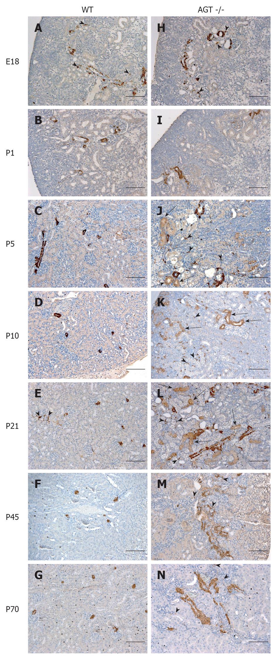
Figure 1 Immunostaining for renin (brown) showing the overall distribution at low magnification in wild type and angiotensinogen deficient mice during embryonic (E18, A and H) and postnatal life (P1, P5, P10, P21, P45 and P70, B-G and I-N).
A: Renin expression in large arteries and arterioles and in pericytes (arrowheads); B: Renin expression in large arteries, arterioles and in the glomerular mesangium; C: Renin expression is still extended in arterioles beyond the juxtaglomerular areas (JGAs); D: Renin expression is mostly restricted to the JGAs; E: Renin expression in JGAs and occasionally in few pericytes (arrowheads); F and G: Renin expression in JGAs; H: Renin expression in large arteries and arterioles and in pericytes (arrowheads) similar to A; I: Renin expression in large arteries and arterioles similar to B; J, L: Renin expression in thickened arteries, arterioles, pericytes (arrowheads) and tubules (arrows); M, N: Renin expression in thickened arteries and arterioles and in fewer pericytes (arrowheads). WT: Wild type; AGT: Angiotensinogen deficient. Bars: 100 μm.
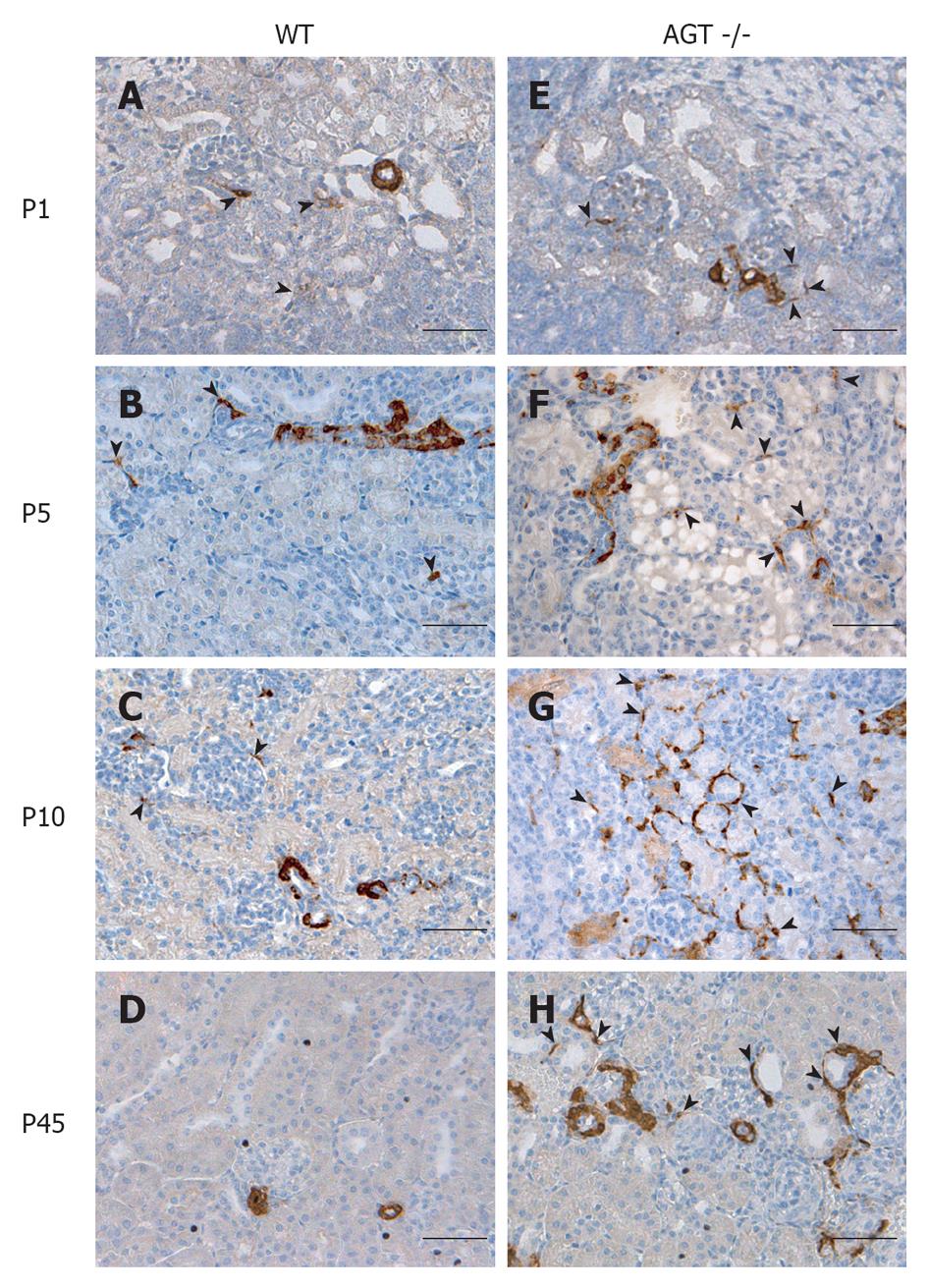
Figure 2 Immunostaining for renin (brown) in wild type (A-D) and angiotensinogen deficient mice (E-H) during postnatal life (P1, P5, P10 and P45) Higher magnification.
A: Renin expression in a transversal section of an arteriole and in isolated pericytes (arrowheads); B: Renin expression in a transversal section of a branching arteriole and in isolated pericytes (arrowheads); C: Renin expression is still present in arterioles beyond the juxtaglomerular areas (JGAs) and in isolated pericytes (arrowheads); D: Renin expression is restricted to the JGAs; E: Renin expression in arterioles and isolated pericytes (arrowheads) similar to A; F: Renin expression in an arteriole beyond the JGA similar to B but with a clear increase in the density of pericytes expressing renin (some marked with arrowheads); G: Peritubular pericytes (some marked with arrowheads) show a marked increase in renin expression; H: Renin expression in enlarged arterioles and in peritubular pericytes (arrowheads). WT: Wild type; AGT: Angiotensinogen deficient. Bars: 50 μm.
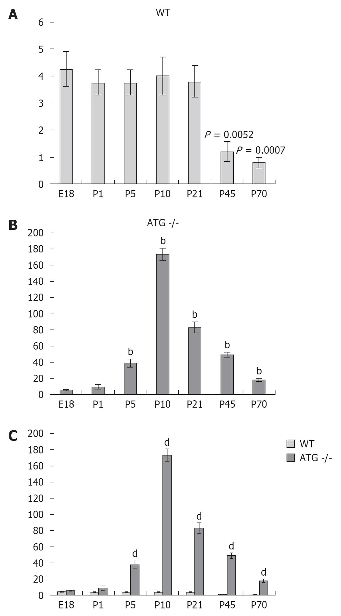
Figure 3 Quantification and comparison of pericytes positive for renin in wild type and angiotensinogen deficient mice.
A: In wild type (WT) mice the density of renin positive pericytes was not significantly different from E18 to P21, but significantly decreased at P45 (P = 0.0052 vs E18) and P70 (P = 0.0007 vs E18); B: Angiotensinogen deficient (AGT) -/- mice showed a significant increase at P5 with a maximum at P10 and a progressive decrease towards P70 (bP≤ 0.01 vs E18); C: Comparison at each individual age showing a significant increase in the number of renin-positive pericytes in AGT -/- from P5 till P70 (dP≤ 0.001 vs WT).
- Citation: Berg AC, Chernavvsky-Sequeira C, Lindsey J, Gomez RA, Sequeira-Lopez MLS. Pericytes synthesize renin. World J Nephrol 2013; 2(1): 11-16
- URL: https://www.wjgnet.com/2220-6124/full/v2/i1/11.htm
- DOI: https://dx.doi.org/10.5527/wjn.v2.i1.11


