Copyright
©The Author(s) 2023.
World J Nephrol. Dec 25, 2023; 12(5): 120-131
Published online Dec 25, 2023. doi: 10.5527/wjn.v12.i5.120
Published online Dec 25, 2023. doi: 10.5527/wjn.v12.i5.120
Figure 1 Various organ involvement of the human body in cryptococcal infection.
UTI: Urinary tract infection; CNS: Central nervous system.
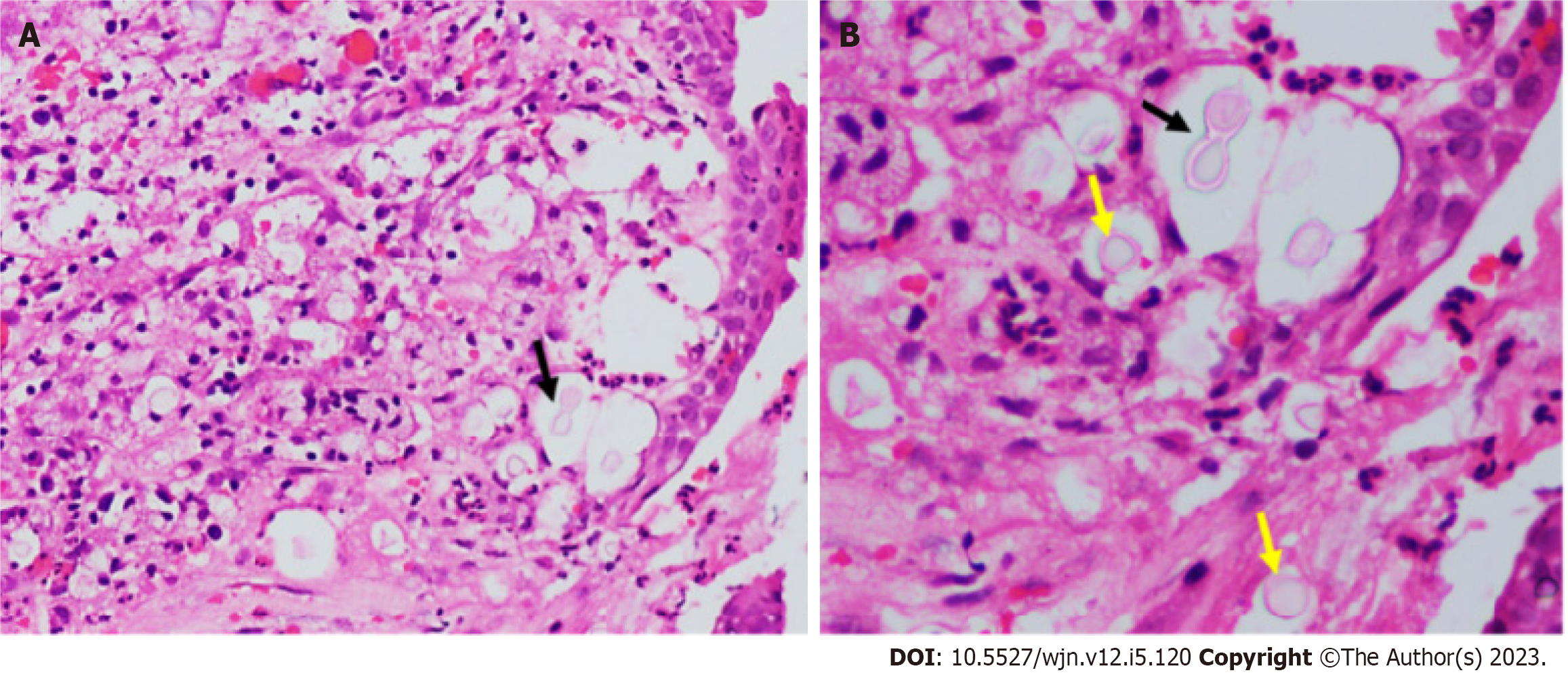
Figure 2 Histopathology of a patient with pulmonary cryptococcosis, hematoxylin and eosin stain and Alcian blue stain.
A: At ×100 magnification; B: At ×200 magnification. Alcian blue-PAS stain atains the yeast forms of cryptococcus. Alcian blue stains the capsule blue colour (black arrow) and PAS stains the cell wall of the yeast magenta colour (yellow arrow).
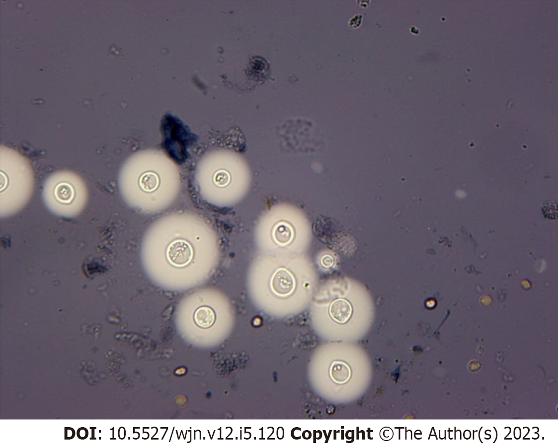
Figure 3 India ink of cryptococcus neoformans.
Endobronchial mucosa shows squamous metaplasia and the sub epithelium shows inflammatory exudates along with variably sized round to oval encapsulated yeast (yellow arrow) with thin walls and narrow based budding (black arrow). A: At ×100 magnification; B: At ×200 magnification.
Figure 4 India ink-stained cryptococcus neoformans.
Figure 5 Therapy for patients with central nervous system disease, disseminated disease or moderate to severe pulmonary involvement.
- Citation: Meena P, Bhargava V, Singh K, sethi J, Prabhakar A, panda S. Cryptococcosis in kidney transplant recipients: Current understanding and practices. World J Nephrol 2023; 12(5): 120-131
- URL: https://www.wjgnet.com/2220-6124/full/v12/i5/120.htm
- DOI: https://dx.doi.org/10.5527/wjn.v12.i5.120


