Copyright
©The Author(s) 2023.
World J Nephrol. Sep 25, 2023; 12(4): 93-103
Published online Sep 25, 2023. doi: 10.5527/wjn.v12.i4.93
Published online Sep 25, 2023. doi: 10.5527/wjn.v12.i4.93
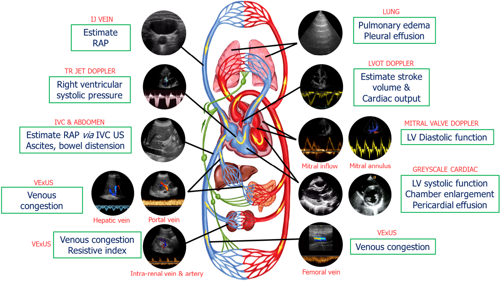
Figure 1 Common sonographic applications used by trained clinicians at the bedside in the evaluation of acute kidney injury.
It is important to evaluate multiple points of the hemodynamic circuit instead of relying on isolated parameters. IJ: Internal jugular; RAP: Right atrial pressure; TR: Tricuspid regurgitation; IVC: Inferior vena cava; US: Ultrasound; VExUS: venous excess ultrasound; LV: Left ventricular; LVOT: Left ventricular outflow tract. Citation: Koratala A, Reisinger N. Point of Care Ultrasound in Cirrhosis-Associated Acute Kidney Injury: Beyond Inferior Vena Cava. Kidney360 2022; 3: 1965-1968. Copyright© 2022 by the American Society of Nephrology (corresponding author’s prior open access publication).
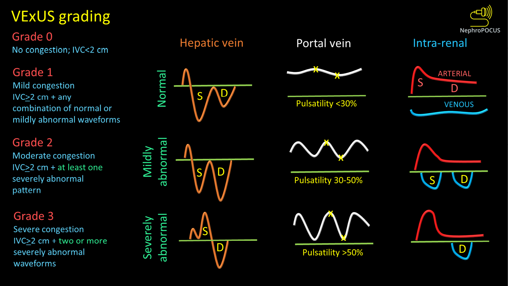
Figure 2 VexUS (Venous Excess Doppler UltraSound) grading system: When the diameter of inferior vena cava is > 2 cm, three grades of congestion are defined based on the severity of abnormalities on hepatic, portal, and renal parenchymal venous Doppler.
Hepatic vein Doppler is considered mildly abnormal when the systolic (S) wave is smaller than the diastolic (D) wave, but still below the baseline; it is considered severely abnormal when the S-wave is reversed. Portal vein Doppler is considered mildly abnormal when the pulsatility is 30% to 50%, and severely abnormal when it is ≥ 50%. Asterisks represent points of pulsatility measurement. Renal parenchymal vein Doppler is mildly abnormal when it is pulsatile with distinct S and D components, and severely abnormal when it is monophasic with D-only pattern. Figure adapted from NephroPOCUS.com with permission (corresponding author’s educational website)-https://nephropocus.com/2021/10/05/vexus-flash-cards/.
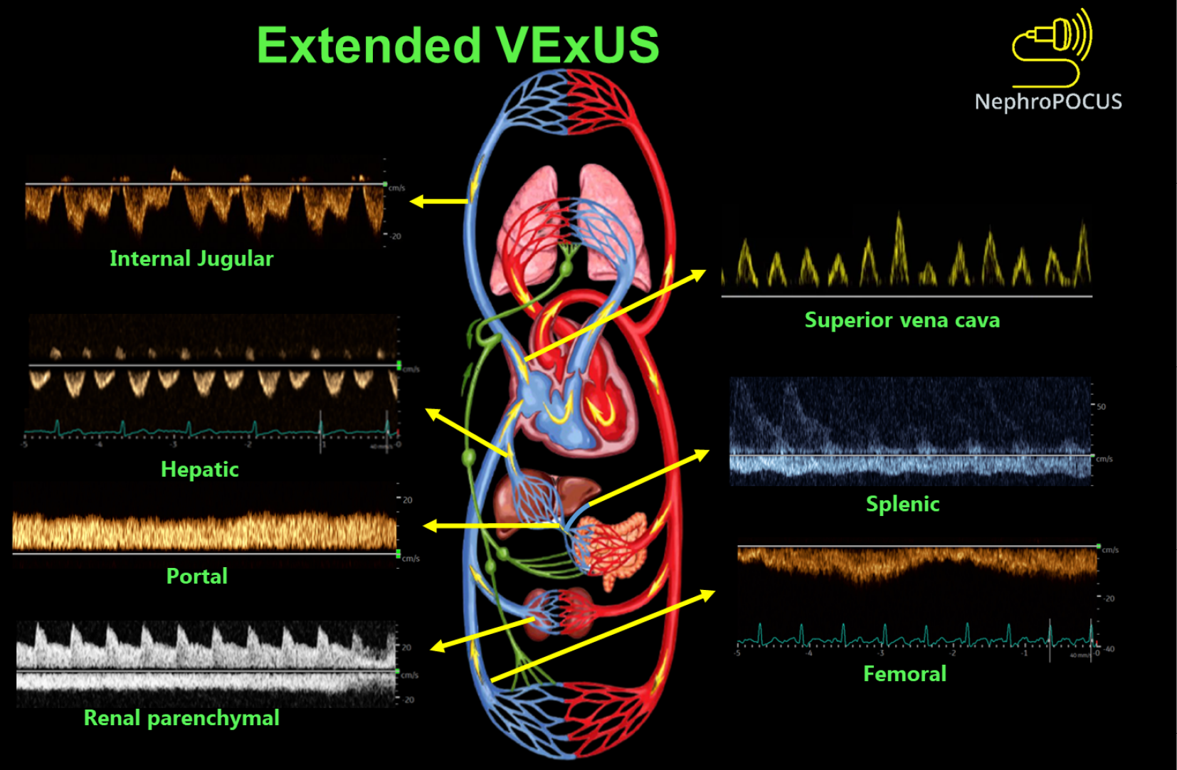
Figure 3 Doppler components of extended VexUS examination.
Figure adapted from NephroPOCUS.com with permission (corresponding author’s educational website)-https://nephropocus.com/2022/11/28/hemodynamic-pocus-in-cirrhosis-think-beyond-the-ivc/.
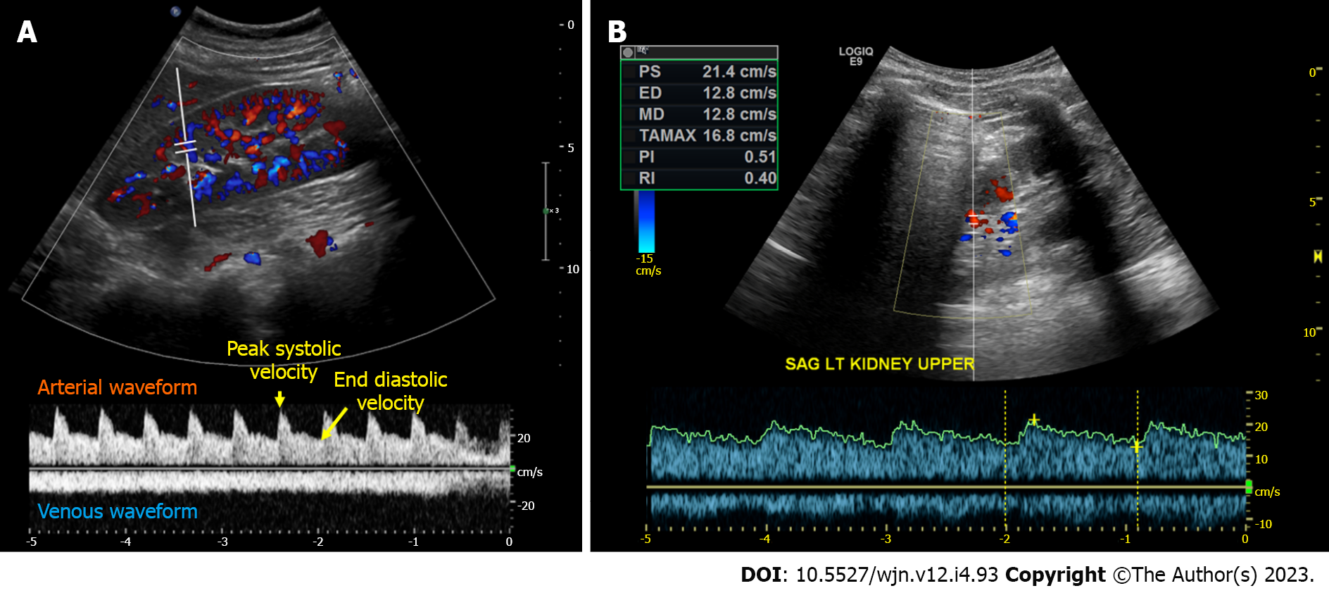
Figure 4 Intrarenal Doppler demonstrating normal waveforms, Tardus Parvus waveform in a case of renal artery stenosis.
A: Normal waveforms; B: Tardus Parvus waveform.
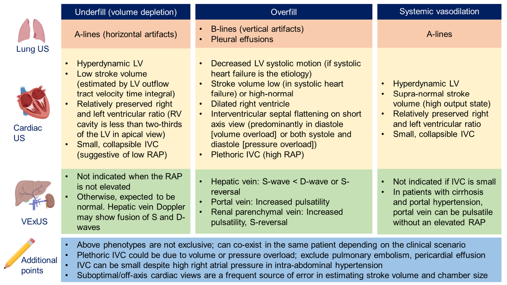
Figure 5 Overview of common ultrasound findings in nephrology-relevant hemodynamic phenotypes.
Systemic vasodilation is frequently seen in patients with liver cirrhosis or early sepsis and renal dysfunction. Underfill phenotype primarily denotes volume depletion. IVC: Inferior vena cava; LV: Left ventricle; RAP: Right atrial pressure; US: Ultrasound; VExUS: Venous excess Doppler ultrasound. Citation: Taleb Abdellah A, Koratala A. Nephrologist-Performed Point-of-Care Ultrasound in Acute Kidney Injury: Beyond Hydronephrosis. Kidney Int Rep 2022; 7: 1428-1432. Copyright© 2022 International Society of Nephrology. Published by Elsevier Inc. (corresponding author’s prior open access publication).
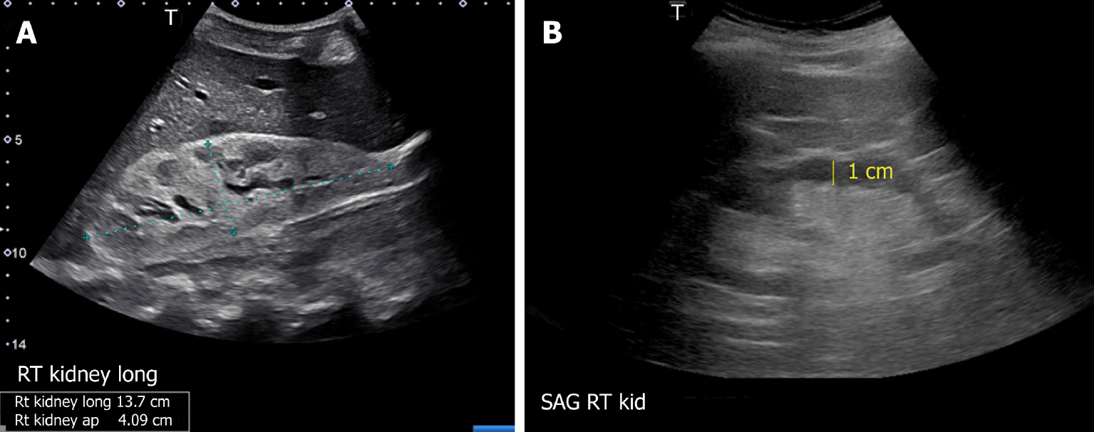
Figure 6 Renal sonogram demonstrating (A) large hyperechogenic kidney in a patient with multiple myeloma and (B) thin parenchyma approximately 1 cm in a patient with chronic kidney disease (normal parenchymal thickness: 1.
5-2 cm). Citation: Koratala A, Bhattacharya D, Kazory A. Point of care renal ultrasonography for the busy nephrologist: A pictorial review. World J Nephrol 2019; 8: 44-58. Copyright© The Author(s) 2019. Published by Baishideng Publishing Group Inc. (corresponding author’s prior open access publication).
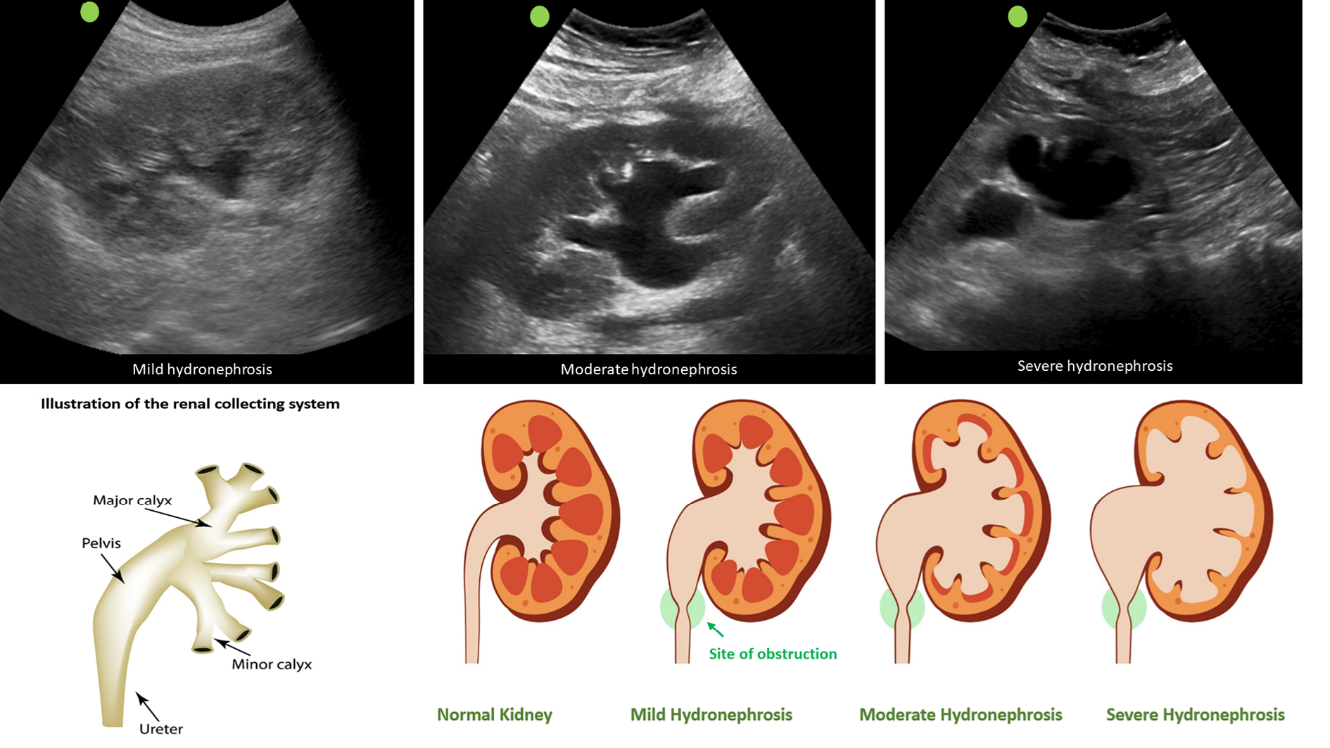
Figure 7 Sonographic appearance of hydronephrosis and its qualitative grading.
Hydronephrosis appears as an anechoic (black) branching structure representing dilated collecting system. Citation: Koratala A. The Ultrasound Mimics of Hydronephrosis. Renal Fellow Network. [cited 18 June 2023]. Available from: https://www.renalfellow.org/2019/05/10/the-ultrasound-mimics-of-hydronephrosis/#:~:text=Prominent%20renal%20vasculature%20and%20vascular,also%20appears%20black%20on%20ultrasound. Copyright© 2019. Published by Renal Fellow Network (corresponding author’s prior open access publication).
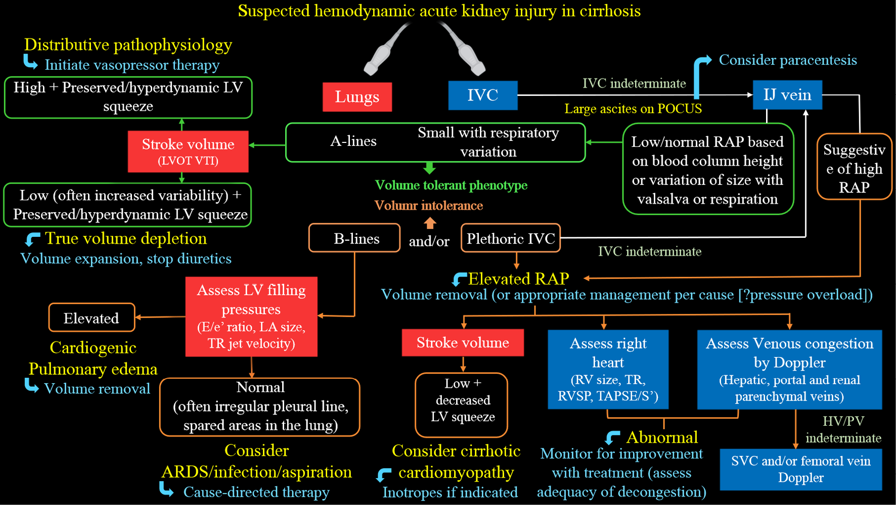
Figure 8 Proposed diagnostic algorithm for approaching acute kidney injury in a patient with cirrhosis.
Citation: Koratala A, Reisinger N. Point of Care Ultrasound in Cirrhosis-Associated Acute Kidney Injury: Beyond Inferior Vena Cava. Kidney360 2022; 3: 1965-1968. Copyright© 2022 by the American Society of Nephrology (corresponding author’s prior open access publication).
- Citation: Batool A, Chaudhry S, Koratala A. Transcending boundaries: Unleashing the potential of multi-organ point-of-care ultrasound in acute kidney injury. World J Nephrol 2023; 12(4): 93-103
- URL: https://www.wjgnet.com/2220-6124/full/v12/i4/93.htm
- DOI: https://dx.doi.org/10.5527/wjn.v12.i4.93


