Copyright
©The Author(s) 2023.
World J Nephrol. May 25, 2023; 12(3): 66-72
Published online May 25, 2023. doi: 10.5527/wjn.v12.i3.66
Published online May 25, 2023. doi: 10.5527/wjn.v12.i3.66
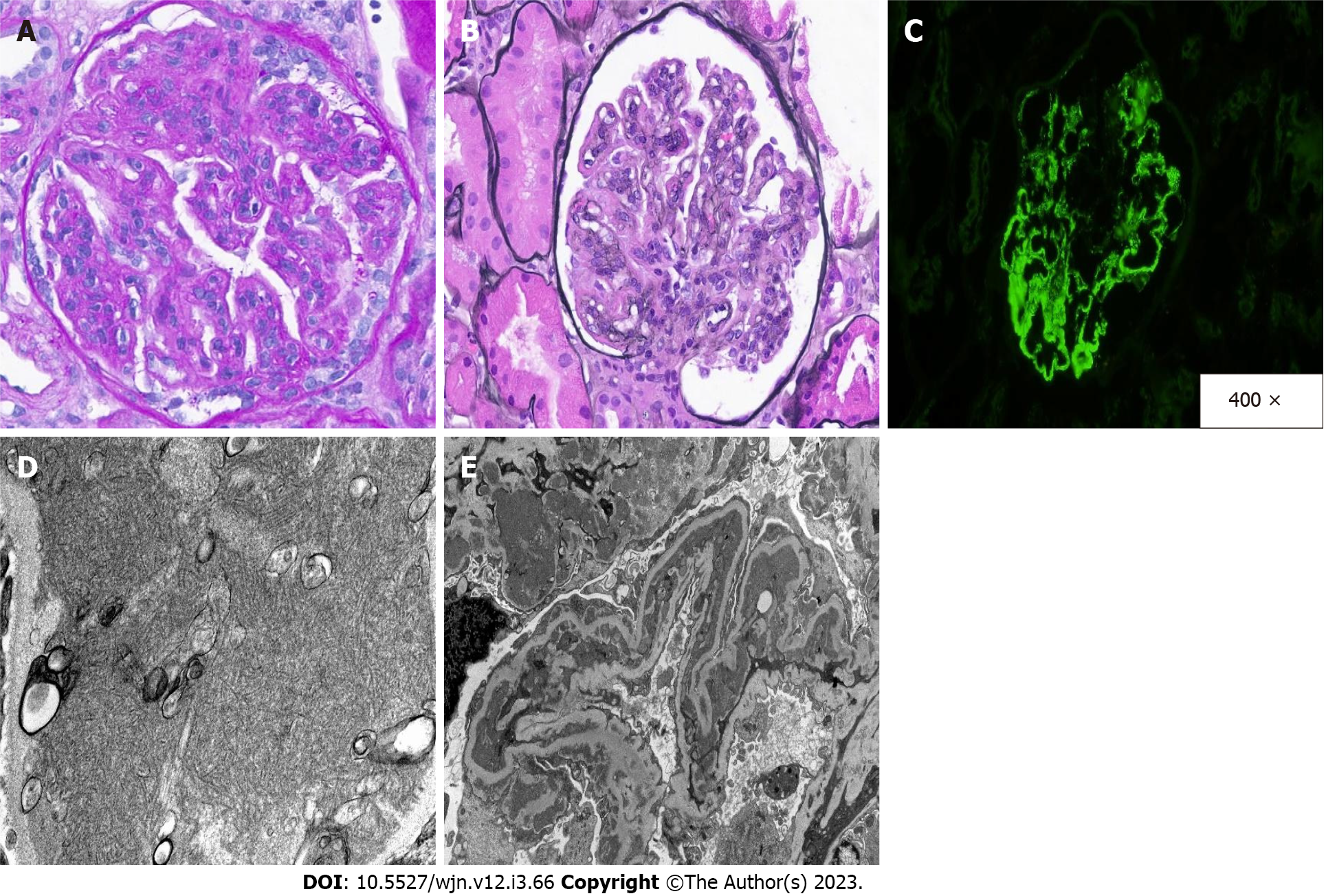
Figure 1 Kidney Biopsy.
A: Light microscopy sections show enlarged glomeruli with lobular accentuation of the capillary loops and both mesangial and endocapillary hypercellularity [periodic acid-Schiff (PAS), 400 ×]; B: Silver-negative, PAS-positive deposits are seen along the capillary loops, resulting in extensive capillary wall remodeling (Jones methenamine silver, 400 ×); C: These deposits show a full-house pattern, highlighting with all immunoglobins and complement components on both standard and pronase immunofluorescence studies (IgG, 400 ×); D and E: On electron microscopy, numerous organized, electron-dense deposits with fibrillary substructure are seen within the mesangial and peripheral capillary loop walls [E: Electron microscopy (EM), 6000 ×], including focal areas with stacked parallel arrays are seen (D: EM, 50000 ×).
- Citation: Lathiya MK, Errabelli P, Mignano S, Cullinan SM. Infection related membranoproliferative glomerulonephritis secondary to anaplasmosis: A case report. World J Nephrol 2023; 12(3): 66-72
- URL: https://www.wjgnet.com/2220-6124/full/v12/i3/66.htm
- DOI: https://dx.doi.org/10.5527/wjn.v12.i3.66


