Copyright
©The Author(s) 2023.
World J Nephrol. Mar 25, 2023; 12(2): 29-39
Published online Mar 25, 2023. doi: 10.5527/wjn.v12.i2.29
Published online Mar 25, 2023. doi: 10.5527/wjn.v12.i2.29
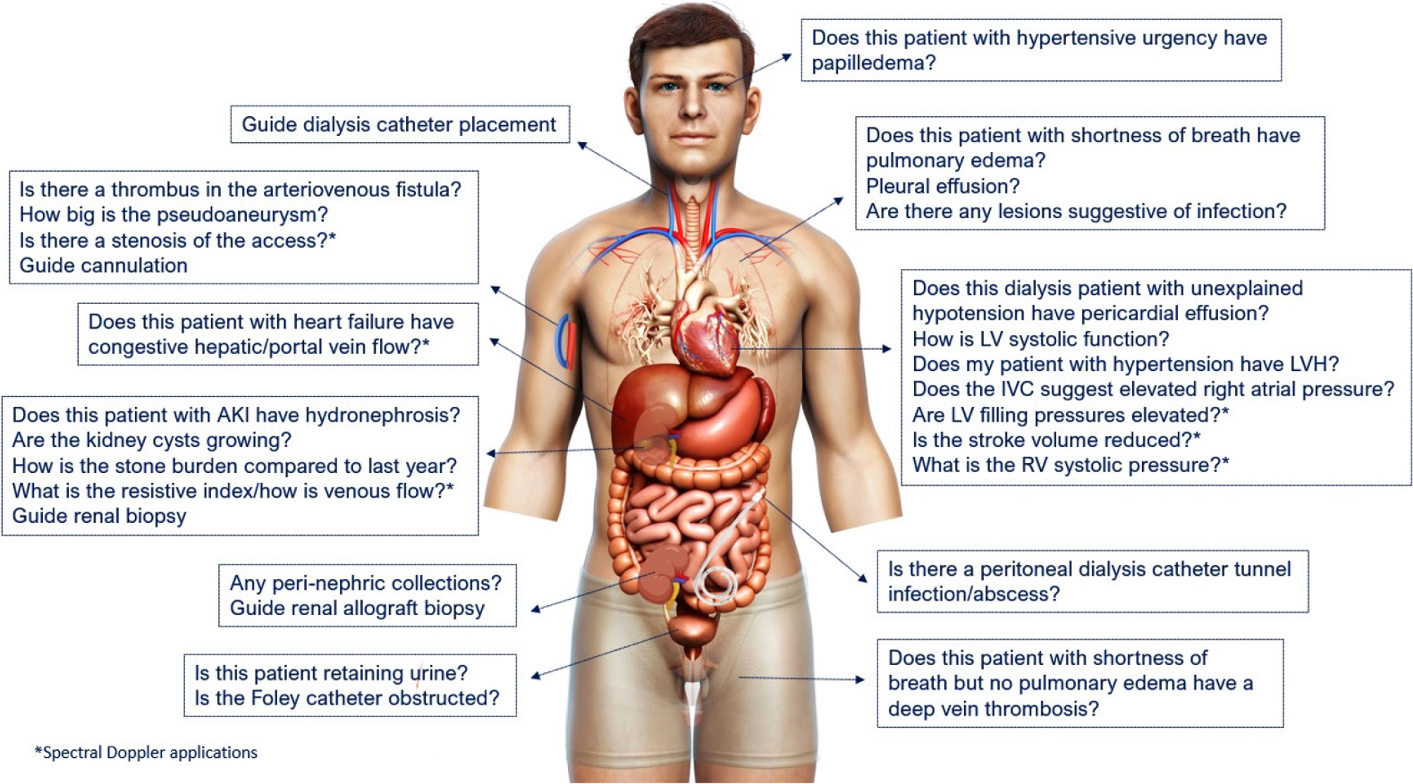
Figure 1 Scope of nephrology-related point of care ultrasonography.
Organ-specific focused questions that can be answered by bedside ultrasonography. Those marked with asterisk (*) indicate advanced sonographic applications requiring a higher operator skill level/additional training. Figure reused from reference # 3 with kind permission of the American Society of Nephrology.
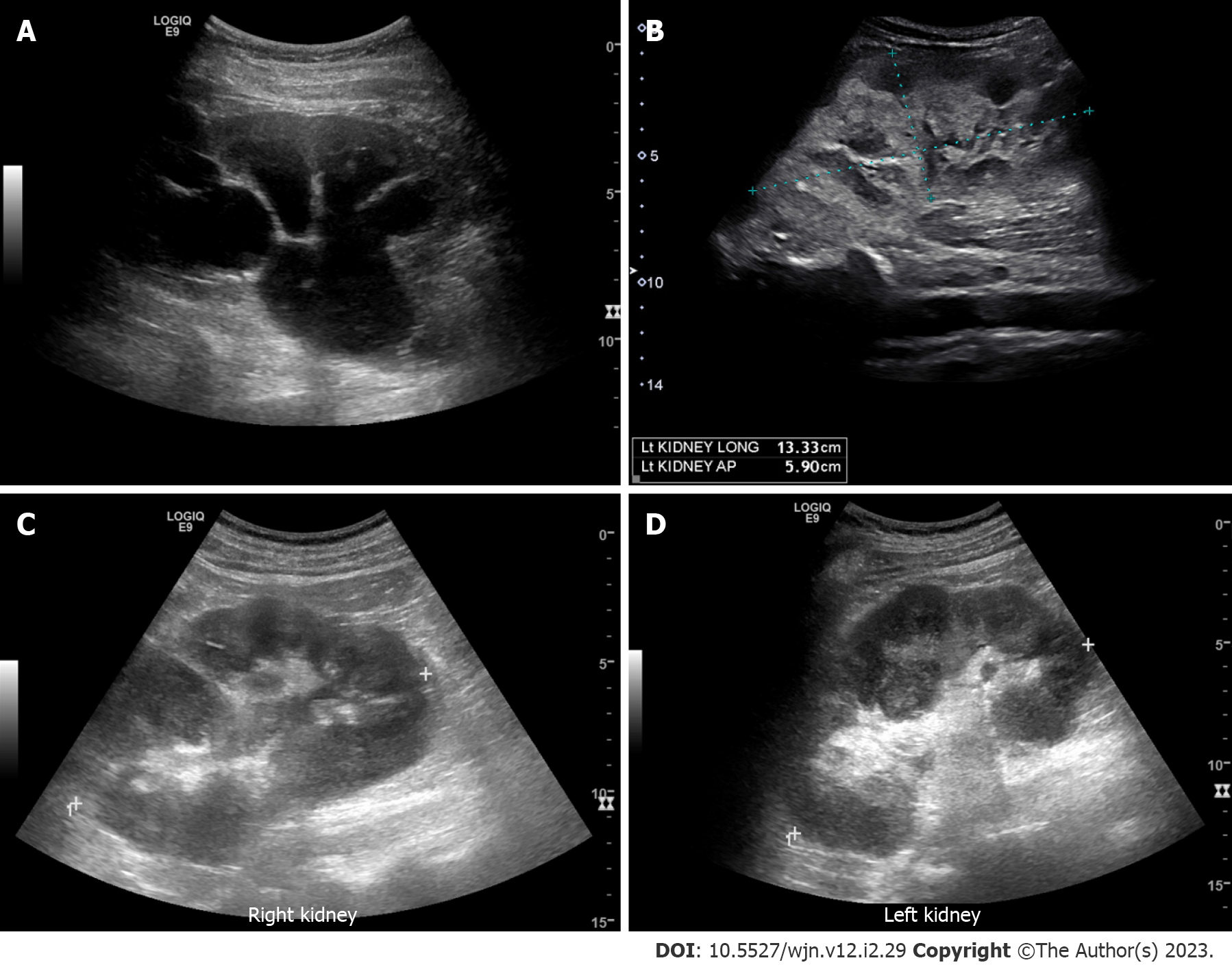
Figure 2 Renal ultrasound images demonstrating.
A: Severe hydronephrosis (branching anechoic area); B: Enlarged kidney with hyperechoic cortex in a patient with myeloma; C and D: Bilateral renal involvement with lymphoma. Note irregular outline and heterogenous parenchyma.
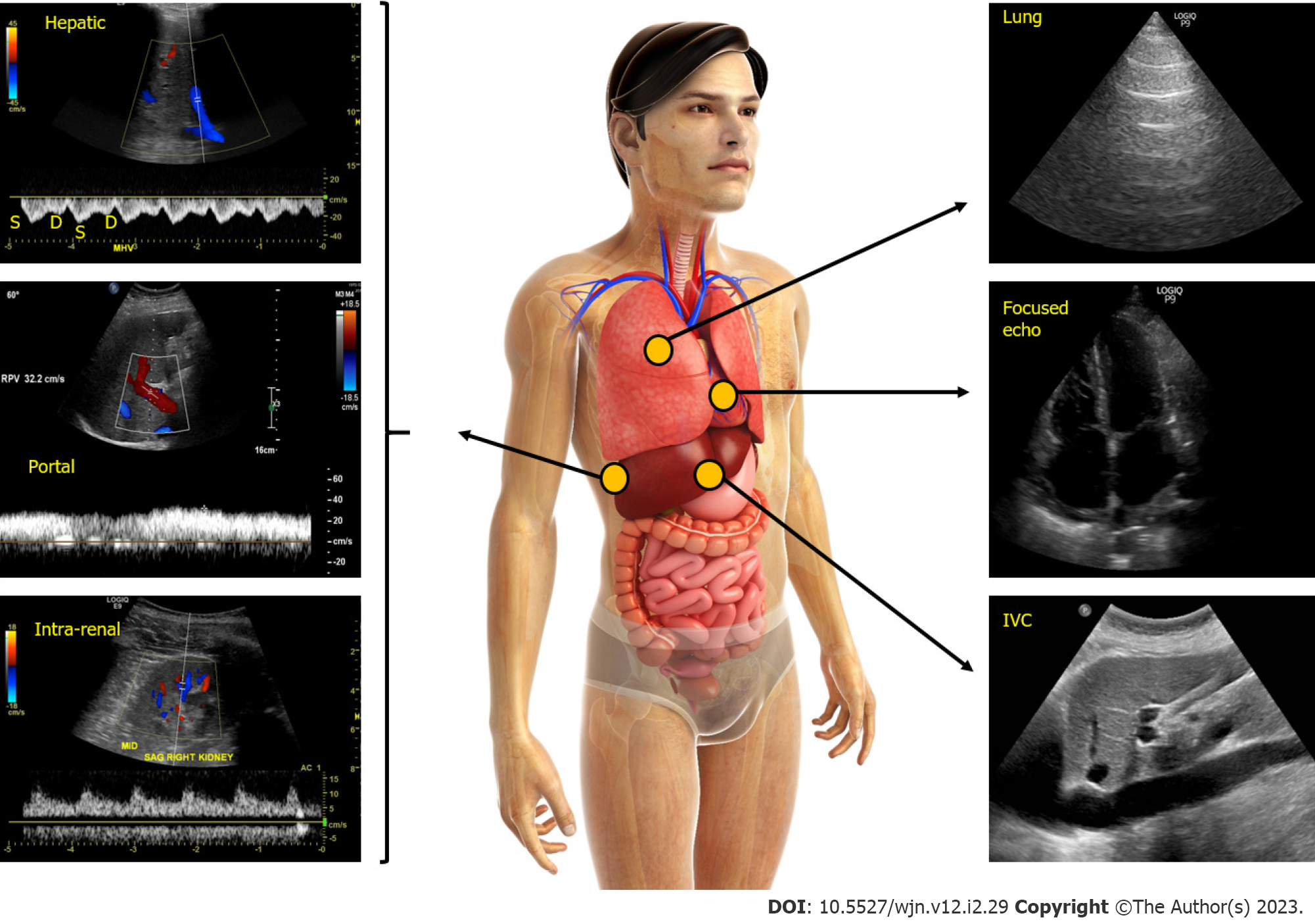
Figure 3 Figure illustrating the integration of multi-point sonographic assessment including focused cardiac, lung and venous Doppler ultrasound.
Normal waveforms are shown. IVC: Inferior vena cava. Adapted from corresponding author’s prior open access publication.
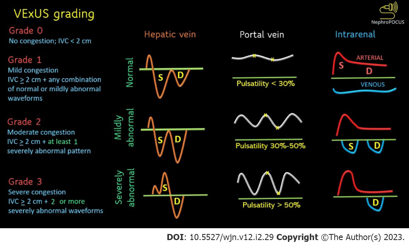
Figure 4 Venous excess ultrasound grading.
When the diameter of inferior vena cava is > 2 cm, three grades of congestion are defined based on the severity of abnormalities on hepatic, portal, and renal parenchymal venous Doppler. Hepatic vein Doppler is considered mildly abnormal when the systolic (S) wave is smaller than the diastolic (D) wave, but still below the baseline; it is considered severely abnormal when the S-wave is reversed. Portal vein Doppler is considered mildly abnormal when the pulsatility is 30% to 50%, and severely abnormal when it is ≥ 50%. Asterisks represent points of pulsatility measurement. Renal parenchymal vein Doppler is mildly abnormal when it is pulsatile with distinct S and D components, and severely abnormal when it is monophasic with D-only pattern. Adapted from NephroPOCUS.com with permission.
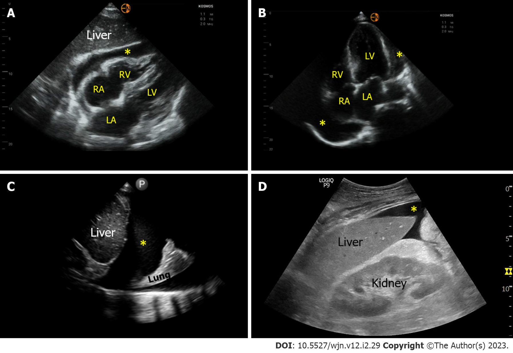
Figure 5 Sonographic images demonstrating various effusions.
A and B: Pericardial effusion (*) as seen on subxiphoid and apical 4-chamber cardiac views respectively; C: Right pleural effusion (*) seen from the lateral scanning window; D: Ascites (*) seen from the right upper quadrant. RA: Right atrium; LA: Left atrium; RV: Right ventricle; LV: Left ventricle.
- Citation: Bhasin-Chhabra B, Koratala A. Point of care ultrasonography in onco-nephrology: A stride toward better physical examination. World J Nephrol 2023; 12(2): 29-39
- URL: https://www.wjgnet.com/2220-6124/full/v12/i2/29.htm
- DOI: https://dx.doi.org/10.5527/wjn.v12.i2.29


