Copyright
©The Author(s) 2022.
World J Nephrol. Jan 25, 2022; 11(1): 30-38
Published online Jan 25, 2022. doi: 10.5527/wjn.v11.i1.30
Published online Jan 25, 2022. doi: 10.5527/wjn.v11.i1.30
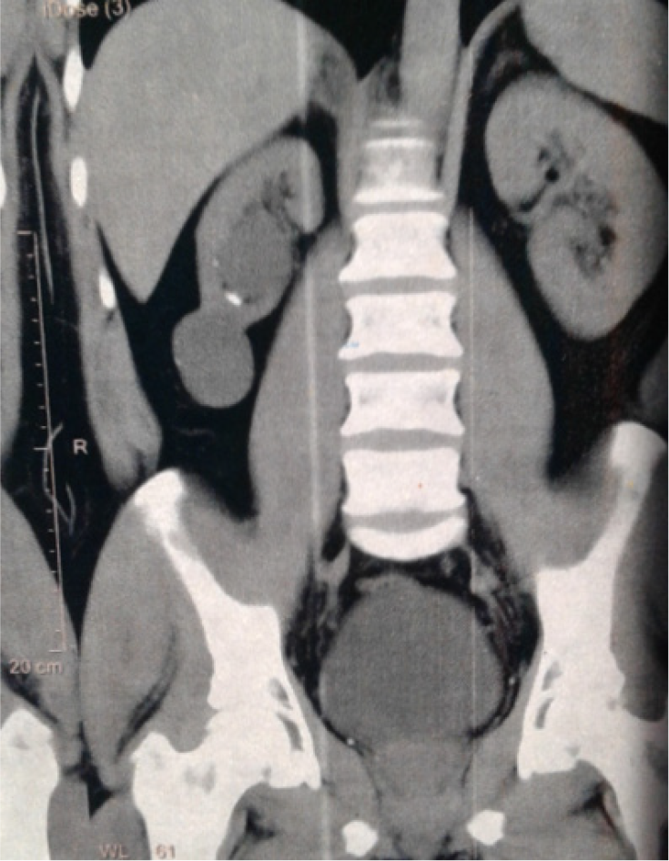
Figure 1 A 44-year-old male patient presented with right loin pain due to right hypoplastic kidney.
A coronal view of non-contrast multi-slice computed tomography of the abdomen and pelvis showing the small-sized right kidney with a smooth outline, two simple cysts at the middle and lower poles, and a very small stone in the lower calyx. This case was managed conservatively.
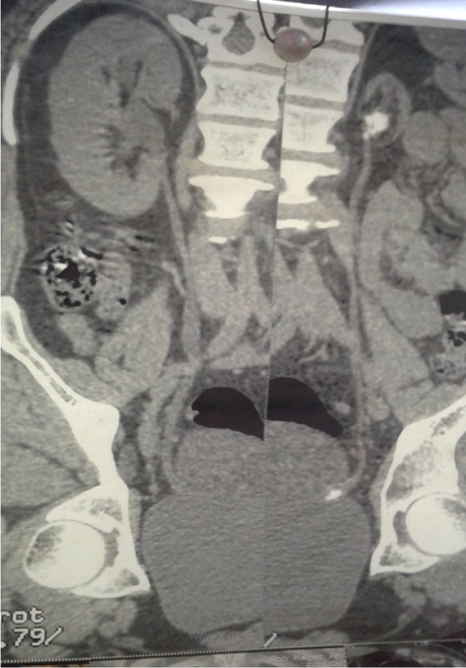
Figure 2 A 43-year-old male patient presented with irritative lower urinary tract symptoms due to a left hypoplastic kidney complicated by stones.
A coronal view of non-contrast multi-slice computed tomography of the abdomen and pelvis showing the severely diminutive left kidney with non-obstructing stones in the renal pelvis and left intramural ureter. This case was managed by left ureteroscopy and nephrectomy.
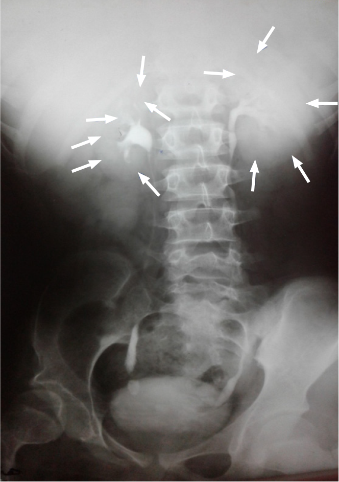
Figure 3 A 39-year-old female patient presented with right loin pain due to right hypoplastic kidney.
An intravenous urography film showing the right hypoplastic kidney with preservation of the normal shape of the pelvicalyceal system and fine details of the whole kidney without obstruction, despite the presence of a right lower ureteral stone. Note the difference between the sizes of both kidneys that are outlined by the arrows.
- Citation: Gadelkareem RA, Mohammed N. Unilateral hypoplastic kidney in adults: An experience of a tertiary-level urology center . World J Nephrol 2022; 11(1): 30-38
- URL: https://www.wjgnet.com/2220-6124/full/v11/i1/30.htm
- DOI: https://dx.doi.org/10.5527/wjn.v11.i1.30


