Copyright
©The Author(s) 2016.
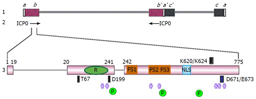
Figure 1 Schematic diagram of infected cell protein 0 gene structure and functional domains.
Line 1: Genome structure of HSV-1; Line 2: Locations of the two inverted copies of ICP0 gene in the HSV-1 genome; Line 3: ICP0 gene structure and domain properties. The amino acid numbers are labeled above the illustration of ICP0 gene. RING finger domain, Proline-rich ND10-FSs and nuclear localization signal are represented by a green oval with “R”, brown squares with “FS” and a blue rectangle with “NLS”, respectively. The binding sites for RNF8 (T67), Cyclin D3 (D199), USP7 (K620/K624), and CoREST (D671/E673) are represented by the dark blue boxes above or beneath the ICP0 gene. The positions of the seven SLSs are represented by lavender hexagons with “S” in the center. The positions of the three phosphorylation clusters are represented by dark green circles with “P” in the center. ICP0: Infected cell protein 0; HSV-1: Herpes simplex virus 1; RING: Really interesting new gene; ND10: Nuclear domains 10; NLS: Nuclear localization signal; USP7: Ubiquitin-specific protease 7; SLSs: SIM-like sequences.
- Citation: Gu H. Infected cell protein 0 functional domains and their coordination in herpes simplex virus replication. World J Virol 2016; 5(1): 1-13
- URL: https://www.wjgnet.com/2220-3249/full/v5/i1/1.htm
- DOI: https://dx.doi.org/10.5501/wjv.v5.i1.1


