Copyright
©The Author(s) 2015.
World J Virology. Aug 12, 2015; 4(3): 156-168
Published online Aug 12, 2015. doi: 10.5501/wjv.v4.i3.156
Published online Aug 12, 2015. doi: 10.5501/wjv.v4.i3.156
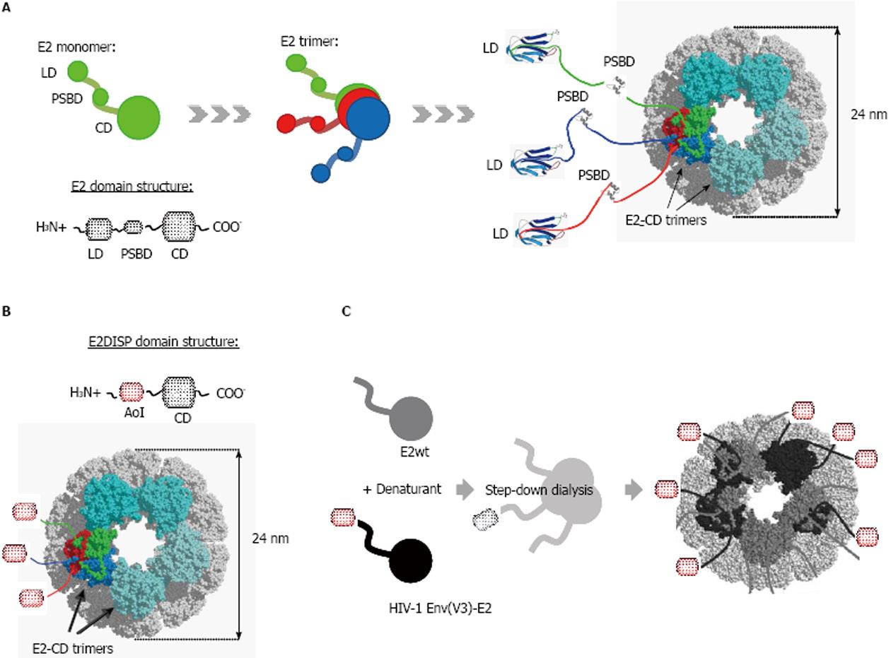
Figure 1 E2 acetyltransferase component from Geobacillus stearothermophilus pyruvate dehydrogenase complex.
A: Schematic illustration of the native E2 chain with: lypoil domain (LD), peripheral subunit-binding domain (PSBD), and catalytic acetyltransferase core domain (CD). E2 CD forms trimers that assemble to generate a pentagonal dodecahedral scaffold(60-mer) with icosahedral symmetry. Trimers are in cyan with monomers of one trimer shown in green, red and blue; B: E2 core from E2DISP acetyltransferase system displaying an antigen of interest (AoI) N-terminally fused to CD; C: Schematic illustration of in vitro refolding of insoluble E2 displaying HIV-1 Envelope V3 in presence of E2 wild-type (E2wt).
- Citation: Trovato M, Berardinis PD. Novel antigen delivery systems. World J Virology 2015; 4(3): 156-168
- URL: https://www.wjgnet.com/2220-3249/full/v4/i3/156.htm
- DOI: https://dx.doi.org/10.5501/wjv.v4.i3.156


