Copyright
©2014 Baishideng Publishing Group Co.
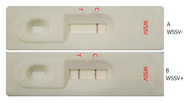
Figure 1 White spot syndrome virus immunochromatographic test strip.
Gill homogenates from (A) uninfected P. monodon showing a negative result with only one reddish-purple band at the C line and (B) WSSV-infected P. monodon showing a positive result with two reddish purple bands at the T line and C line. T: Test line; C: Control line. WSSV: White spot syndrome virus.
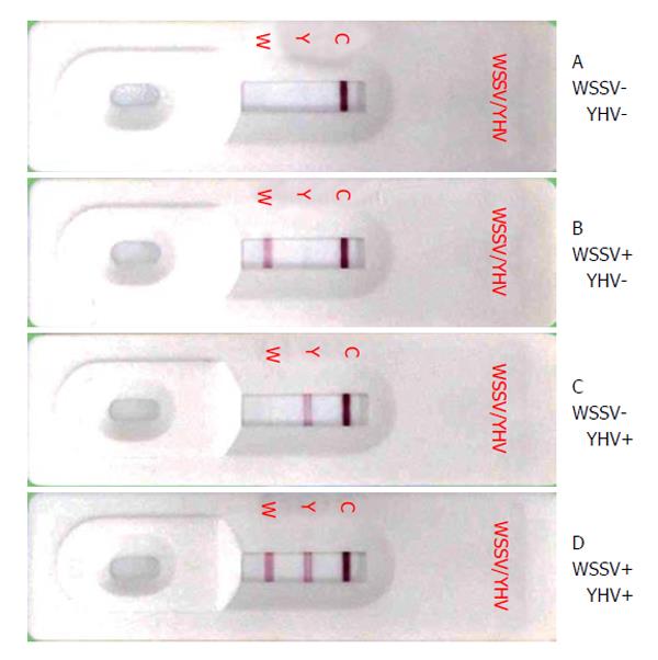
Figure 2 White spot syndrome virus and yellow head virus Dual test strip results.
Pleopod homogenates samples from (A) uninfected P. vannamei, (B) WSSV infected P. vannamei, (C) YHV infected P. vannamei or (D) a combination of B and C were applied to the test strip. W: The test line for WSSV, Y: The test line for YHV and C: The control line. WSSV: White spot syndrome virus; YHV: Yellow head virus.
Figure 3 Infectious myonecrosis virus test strip results.
Shrimp muscle homogenate samples from (A) an uninfected P. vannamei showing a negative result with only one reddish-purple band at the C line and (B) an IMNV infected P. vannamei showing a positive result with two reddish purple bands. IMNV: Infectious myonecrosis virus.
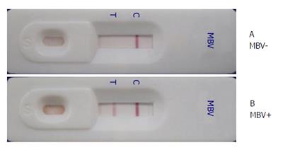
Figure 4 Penaeus monodon nucleopolyhedrovirus immunochromatographic strip test results.
Homogenates of (A) uninfected P. monodon postlarvae showing a negative result with only one band at the C line and (B) PemoNPV-infected P. monodon postlarvae displaying a positive result. PemoNPV: Penaeus monodon nucleopolyhedrovirus; MBV: Monodon baculovirus.
- Citation: Chaivisuthangkura P, Longyant S, Sithigorngul P. Immunological-based assays for specific detection of shrimp viruses. World J Virol 2014; 3(1): 1-10
- URL: https://www.wjgnet.com/2220-3249/full/v3/i1/1.htm
- DOI: https://dx.doi.org/10.5501/wjv.v3.i1.1


