Copyright
©The Author(s) 2024.
World J Virol. Dec 25, 2024; 13(4): 95986
Published online Dec 25, 2024. doi: 10.5501/wjv.v13.i4.95986
Published online Dec 25, 2024. doi: 10.5501/wjv.v13.i4.95986
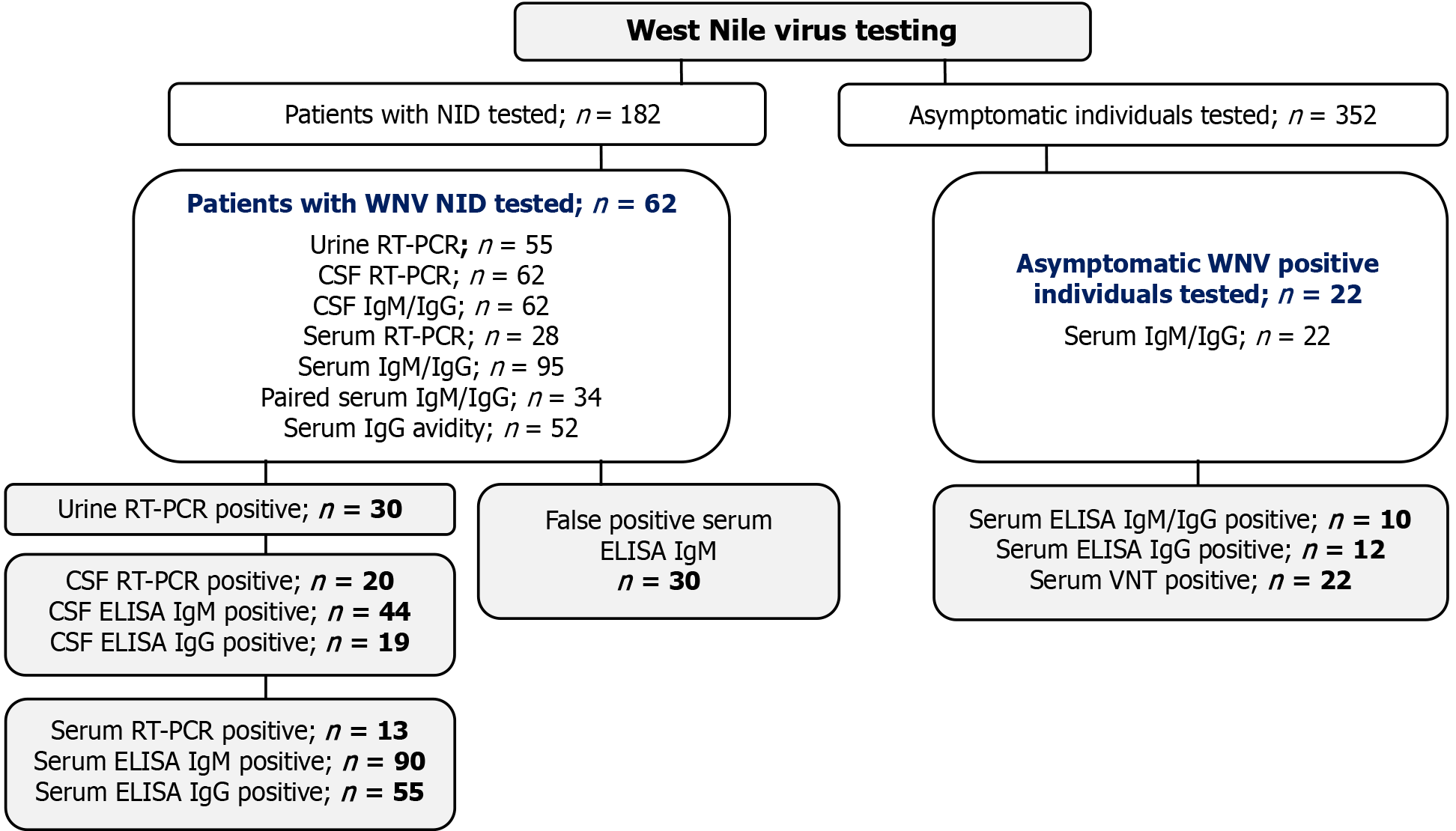
Figure 1 Selection of patients with West Nile virus infection and clinical samples.
Samples in gray shadowed boxes were analyzed in the study. CSF: Cerebrospinal fluid; IgG: Immunoglobulin G; IgM: Immunoglobulin M; NID: Neuroinvasive disease; RT-PCR: Reverse transcription-polymerase chain reaction; TBEV: Tick-borne encephalitis virus; USUV: Usutu virus; WNV: West Nile virus.
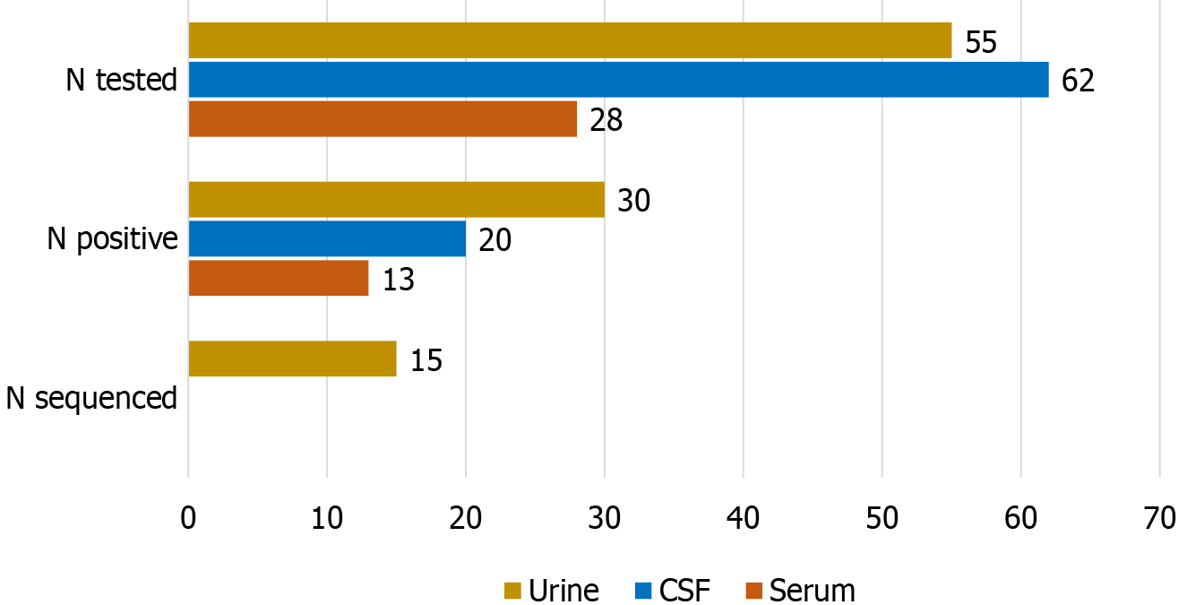
Figure 2 Detection rate of West Nile virus RNA by reverse transcription-polymerase chain reaction.
CSF: Cerebrospinal fluid; N: Total sample number.
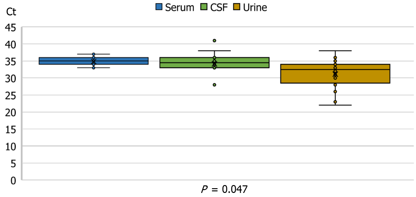
Figure 3 West Nile virus reverse transcription-polymerase chain reaction cycle threshold values in different clinical samples.
Plots represent medians with interquartile ranges, inner and outlier points. CSF: Cerebrospinal fluid; Ct: Cycle threshold.
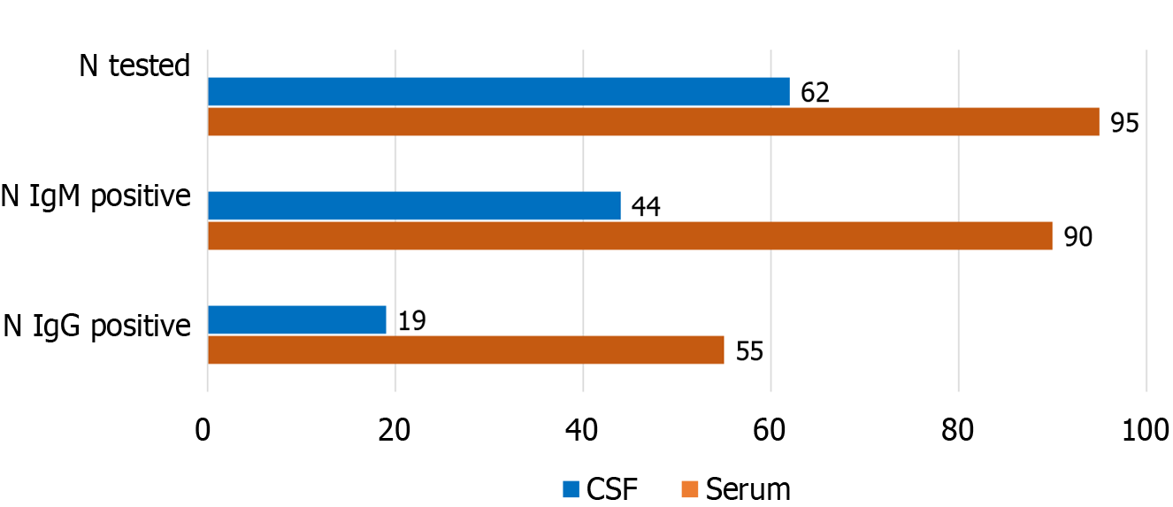
Figure 4 West Nile virus immunoglobulin M and immunoglobulin G detection rates in serum and cerebrospinal fluid.
CSF: Cerebrospinal fluid; Ig: Immunoglobulin; N: Total sample number.
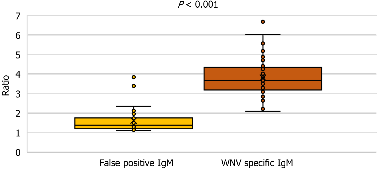
Figure 5 Immunoglobulin M antibody levels in patients with confirmed West Nile virus infection and false positive samples.
Plots represent medians with interquartile ranges, inner and outlier points. IgM: Immunoglobulin M; WNV: West Nile virus.
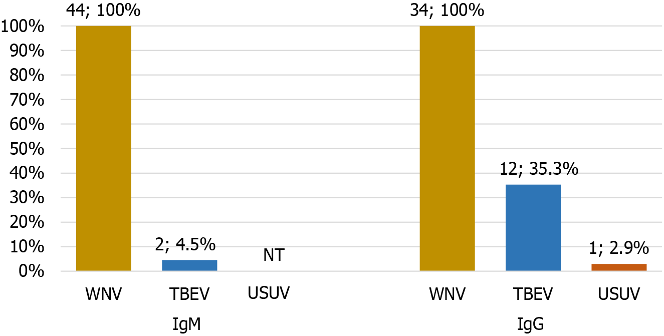
Figure 6 Cross-reactive flavivirus immunoglobulin M immunoglobulin G antibodies detected in patients with West Nile virus infections.
IgG: Immunoglobulin G; IgM: Immunoglobulin M; NT: Not tested; TBEV: Tick-borne encephalitis virus; USUV: Usutu virus; WNV: West Nile virus.
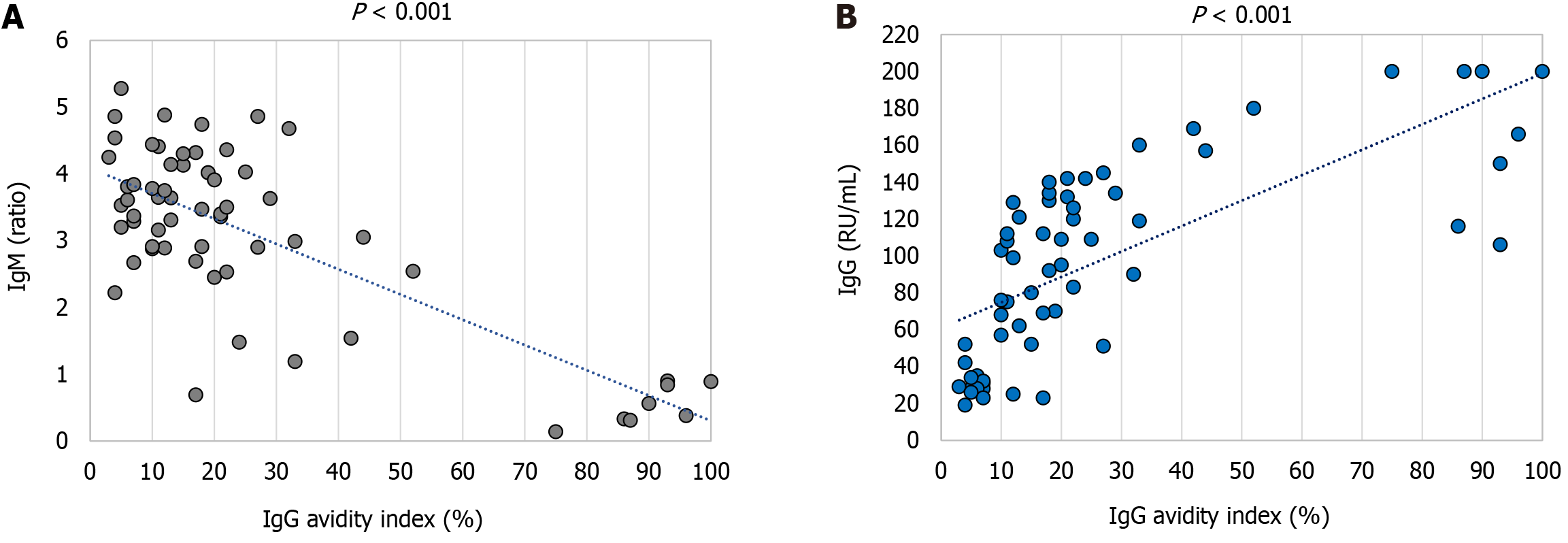
Figure 7 Correlation of immunoglobulin G avidity.
A: Immunoglobulin M antibody levels; B: Immunoglobulin G antibody levels. Blue dotted lines represent trendlines. IgG: Immunoglobulin G; IgM: Immunoglobulin M.
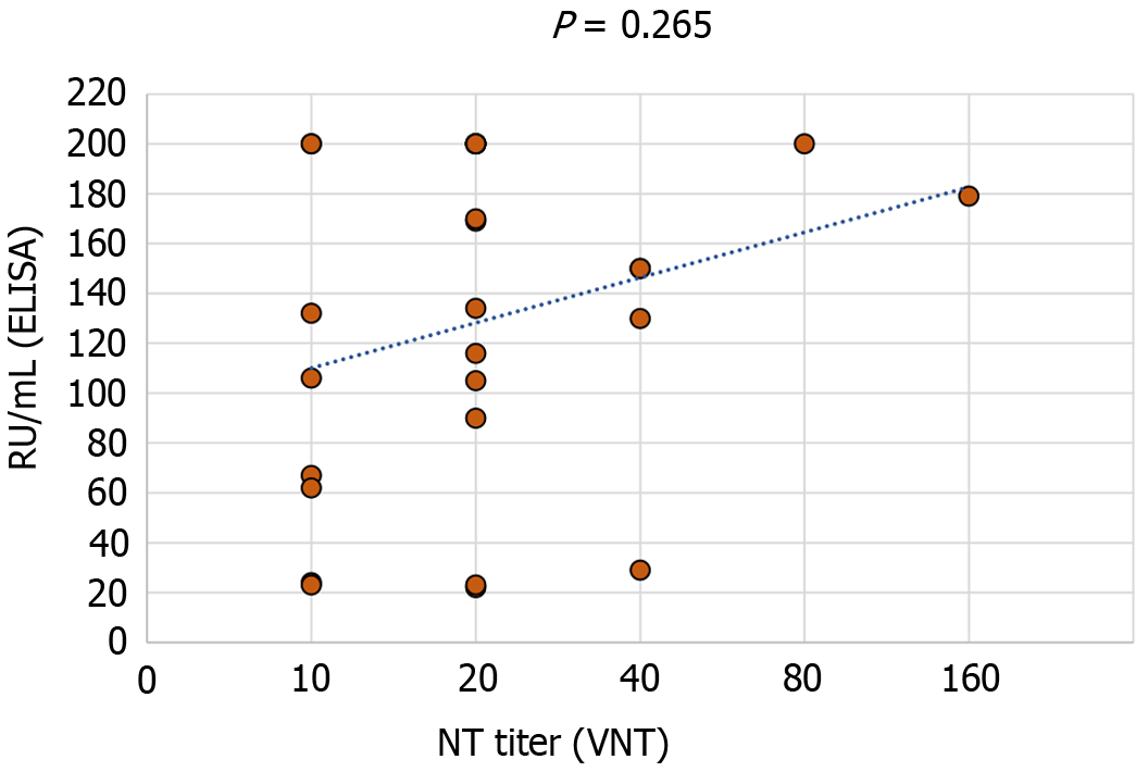
Figure 8 Correlation of immunoglobulin G antibody levels (ELISA) and NT titer (Virus neutralization test).
Blue dotted line represents a trendline.
- Citation: Vilibic-Cavlek T, Bogdanic M, Savic V, Hruskar Z, Barbic L, Stevanovic V, Antolasic L, Milasincic L, Sabadi D, Miletic G, Coric I, Mrzljak A, Listes E, Savini G. Diagnosis of West Nile virus infections: Evaluation of different laboratory methods. World J Virol 2024; 13(4): 95986
- URL: https://www.wjgnet.com/2220-3249/full/v13/i4/95986.htm
- DOI: https://dx.doi.org/10.5501/wjv.v13.i4.95986


