Copyright
©The Author(s) 2017.
World J Transplant. Jun 24, 2017; 7(3): 179-192
Published online Jun 24, 2017. doi: 10.5500/wjt.v7.i3.179
Published online Jun 24, 2017. doi: 10.5500/wjt.v7.i3.179
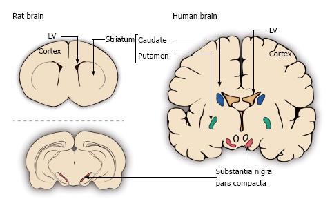
Figure 3 Schematic representation of different sites in the rat and human brains used for grafting in Parkinson’s disease.
The depicted grafting sites include the lateral ventricles (LV), the striatum (in rat) or caudate nucleus and putamen (in human) and the substantia nigra pars compacta. The above schemes are coronal sections of the rat striatum and human caudate (blue) and putamen (green) together with the substantia nigra pars compacta (red). The scheme below is a coronal section at the level of the rat substantia nigra pars compacta (red).
- Citation: Boronat-García A, Guerra-Crespo M, Drucker-Colín R. Historical perspective of cell transplantation in Parkinson’s disease. World J Transplant 2017; 7(3): 179-192
- URL: https://www.wjgnet.com/2220-3230/full/v7/i3/179.htm
- DOI: https://dx.doi.org/10.5500/wjt.v7.i3.179


