Copyright
©The Author(s) 2018.
World J Transplantation. Oct 22, 2018; 8(6): 232-236
Published online Oct 22, 2018. doi: 10.5500/wjt.v8.i6.232
Published online Oct 22, 2018. doi: 10.5500/wjt.v8.i6.232
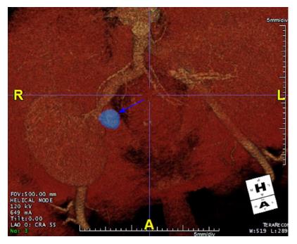
Figure 1 Computerized tomography angiogram showing a 20 mm x 25 mm pseudoaneurysm arising from the aortic patch (blue arrow).
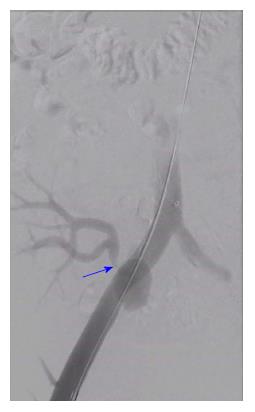
Figure 2 Angiogram showing transplant renal artery stenosis (blue arrow) due to compression caused by the pseudoaneurysm.
Guide wire is present within the right common and external iliac artery.
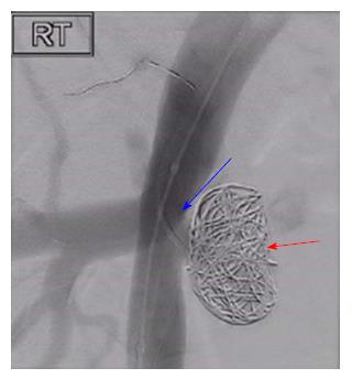
Figure 3 Successful coiling of the pseudoaneurysm (red arrow) and stenting of the transplant renal artery stenosis (blue arrow).
- Citation: Marie Y, Kumar A, Hinchliffe S, Curran S, Brown P, Turner D, Shrestha B. Treatment of transplant renal artery pseudoaneurysm using expandable hydrogel coils: A case report and review of literature. World J Transplantation 2018; 8(6): 232-236
- URL: https://www.wjgnet.com/2220-3230/full/v8/i6/232.htm
- DOI: https://dx.doi.org/10.5500/wjt.v8.i6.232


