Copyright
©The Author(s) 2018.
World J Transplantation. Sep 10, 2018; 8(5): 142-149
Published online Sep 10, 2018. doi: 10.5500/wjt.v8.i5.142
Published online Sep 10, 2018. doi: 10.5500/wjt.v8.i5.142
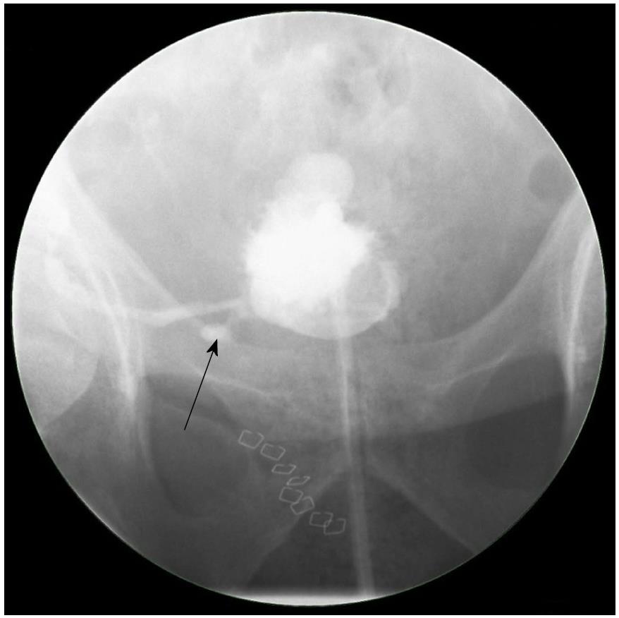
Figure 1 A cystogram showing urinary leak (arrow) at the anastomosis between the newly implanted graft ureter and urinary bladder.
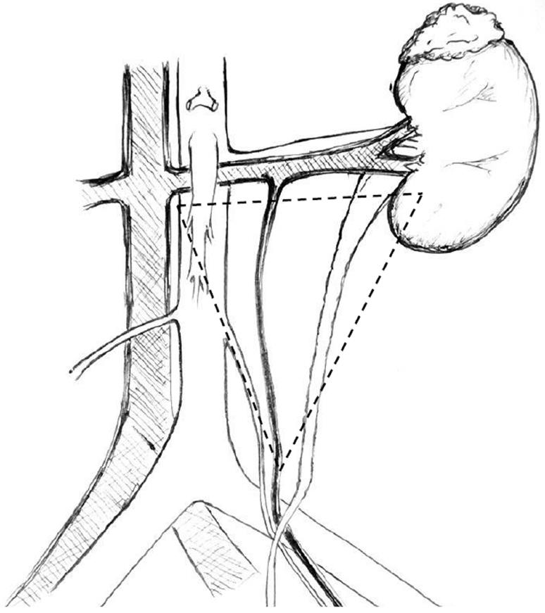
Figure 2 The golden triangle.
Bordered by the lower pole of the kidney on the left, the junction between the renal vein and the inferior vena cava on the right and gonadal vein.
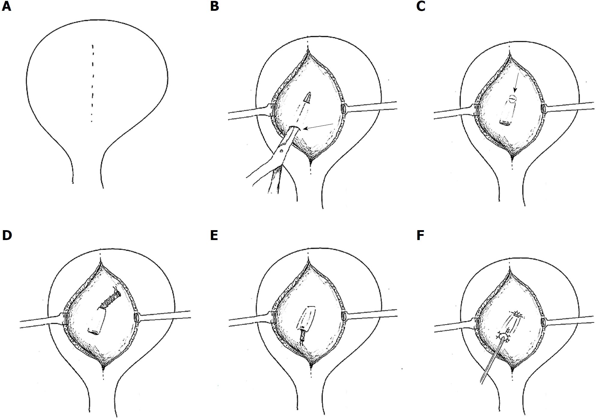
Figure 3 Leadbetter-Politano technique.
A: A longitudinal bladder incision is performed to gain access to the interior of the bladder; B: A second cystotomy is done to introduce the neo-ureter in the bladder. Subsequently, an Overholt is inserted from the second cystotomy and tunnelled close to the bladder wall for about 3 cm; C: A new hiatus is created at the end of the tunnel; D: The neo-ureter is pulled through the mucosal tunnel and the new mucosal hiatus using a free suture as a guide rail; E: Closure of the second cystotomy and then sub-mucosal transposition of distal neo-ureter; F: Fixation of the neo-ureter orifice and closure of the bladder mucosa.
Figure 4 Lich-Gregoir technique.
A: Bladder wall incision through the detrusor muscle is performed, leaving a very thin layer of muscle and uroepithelium unbreached; B: The distal part is completely incised to create a neo-ureter-bladder anastomosis; C: Suturing of the neo-ureter is performed via the same access used to introduce it into the bladder; D: The ureter is positioned in the groove and in direct contact to the uroepithelium, followed by closure of the muscle over the ureter while carefully avoiding constriction of the neo-ureter.
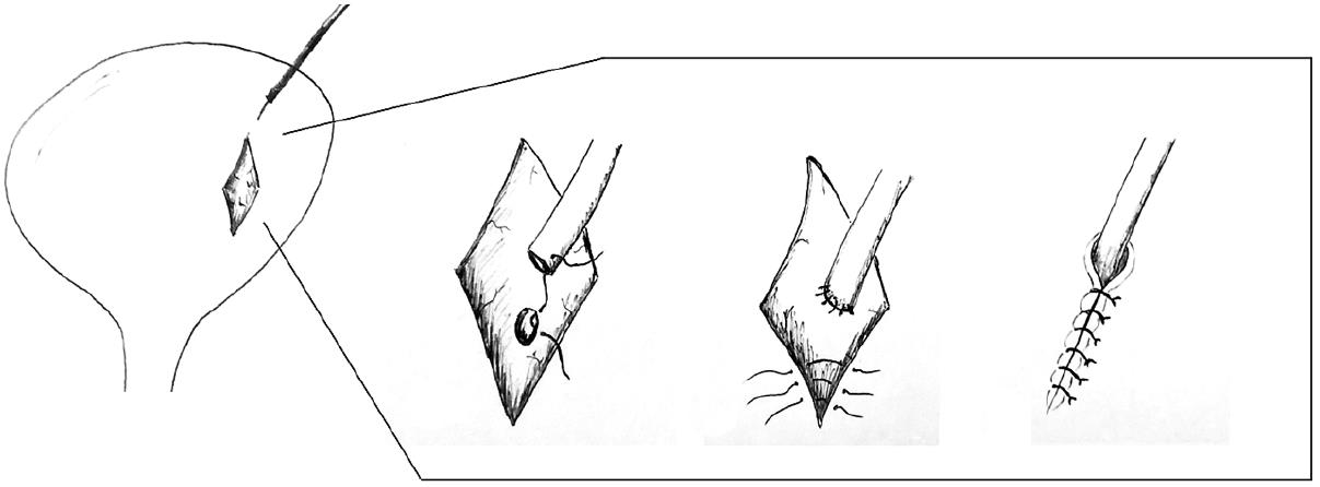
Figure 5 Taguchi technique.
A suture is positioned at the distal end of the neo-ureter and subsequently introduced in the bladder via a cystotomy. The neo-ureter is later fixed to the bladder wall by bringing the suture out through the bladder wall and closed.
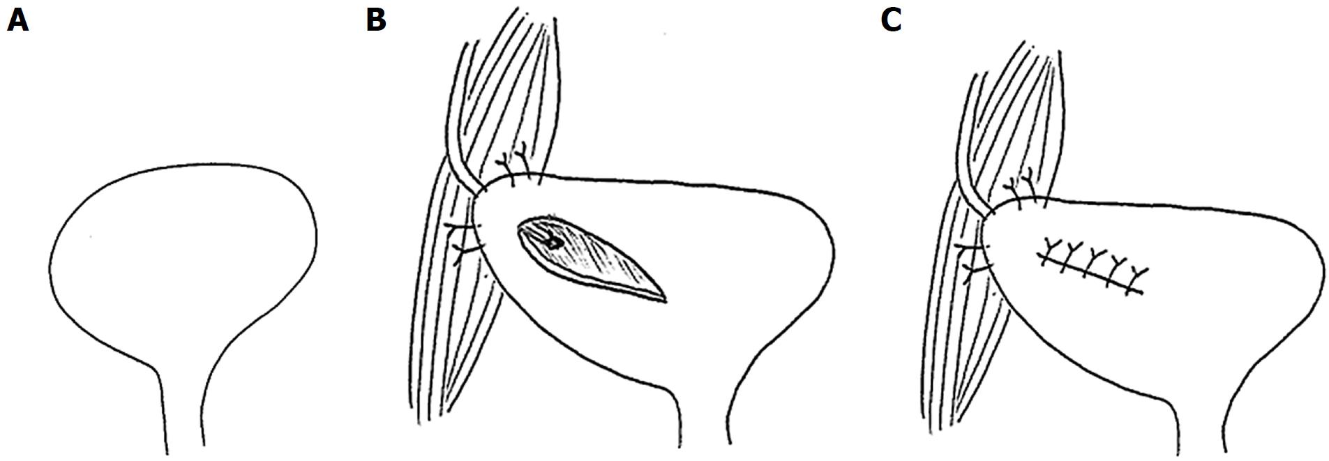
Figure 6 Psoas hitch.
A: A psoas hitch procedure is used to bridge the gap between the urinary bladder and a short ureter; B: Mobilization of the urinary bladder is achieved by dissecting the attachments of the urinary bladder, which is subsequently hitched to the Psoas muscle; C: Ureter implantation is performed via a transverse incision, which is later closed.
Figure 7 Boari flap.
A: A Boari flap is used when a Psoas hitch is not enough to bridge the gap between the bladder and a short ureter to allow for a tension-free anastomosis. A U-shaped flap composed of all tissue layers is created. The base should be proportional to the length of the flap to avoid ischemia; B: The ureter is implanted to the apex of the flap via end-to-end anastomosis or a sub-mucosal tunnel; C: The bladder incision together with the flap are subsequently closed.
- Citation: Buttigieg J, Agius-Anastasi A, Sharma A, Halawa A. Early urological complications after kidney transplantation: An overview. World J Transplantation 2018; 8(5): 142-149
- URL: https://www.wjgnet.com/2220-3230/full/v8/i5/142.htm
- DOI: https://dx.doi.org/10.5500/wjt.v8.i5.142


