Copyright
©The Author(s) 2017.
World J Transplant. Dec 24, 2017; 7(6): 359-363
Published online Dec 24, 2017. doi: 10.5500/wjt.v7.i6.359
Published online Dec 24, 2017. doi: 10.5500/wjt.v7.i6.359
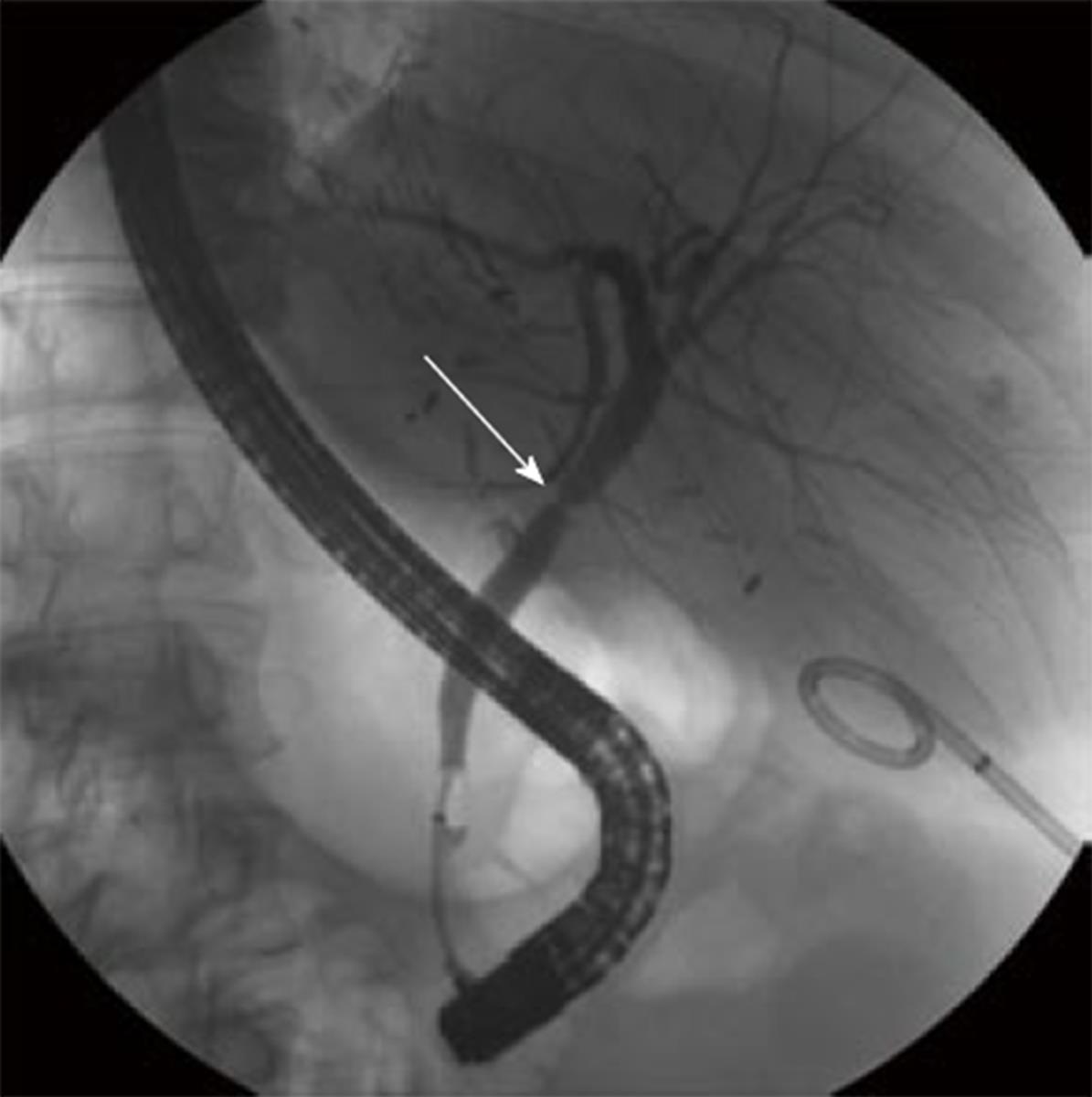
Figure 1 Endoscopic retrograde cholangiopancreatography which showed biliary stricture.
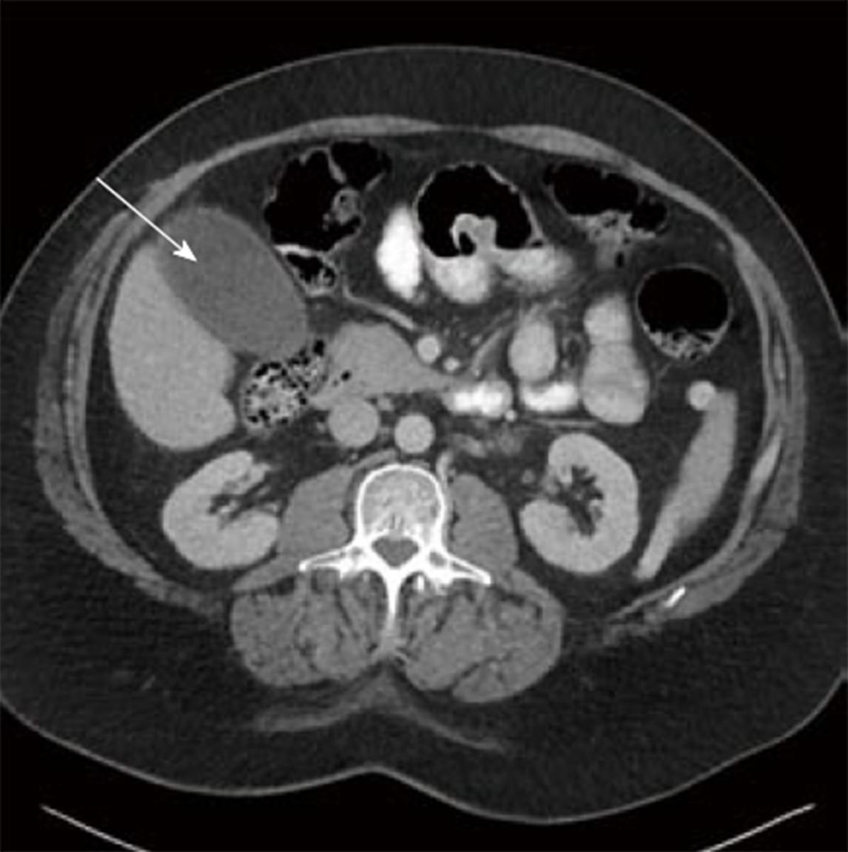
Figure 2 Collection inferior to segment 5 in the donor gall bladder fossa mimicking a gallbladder.
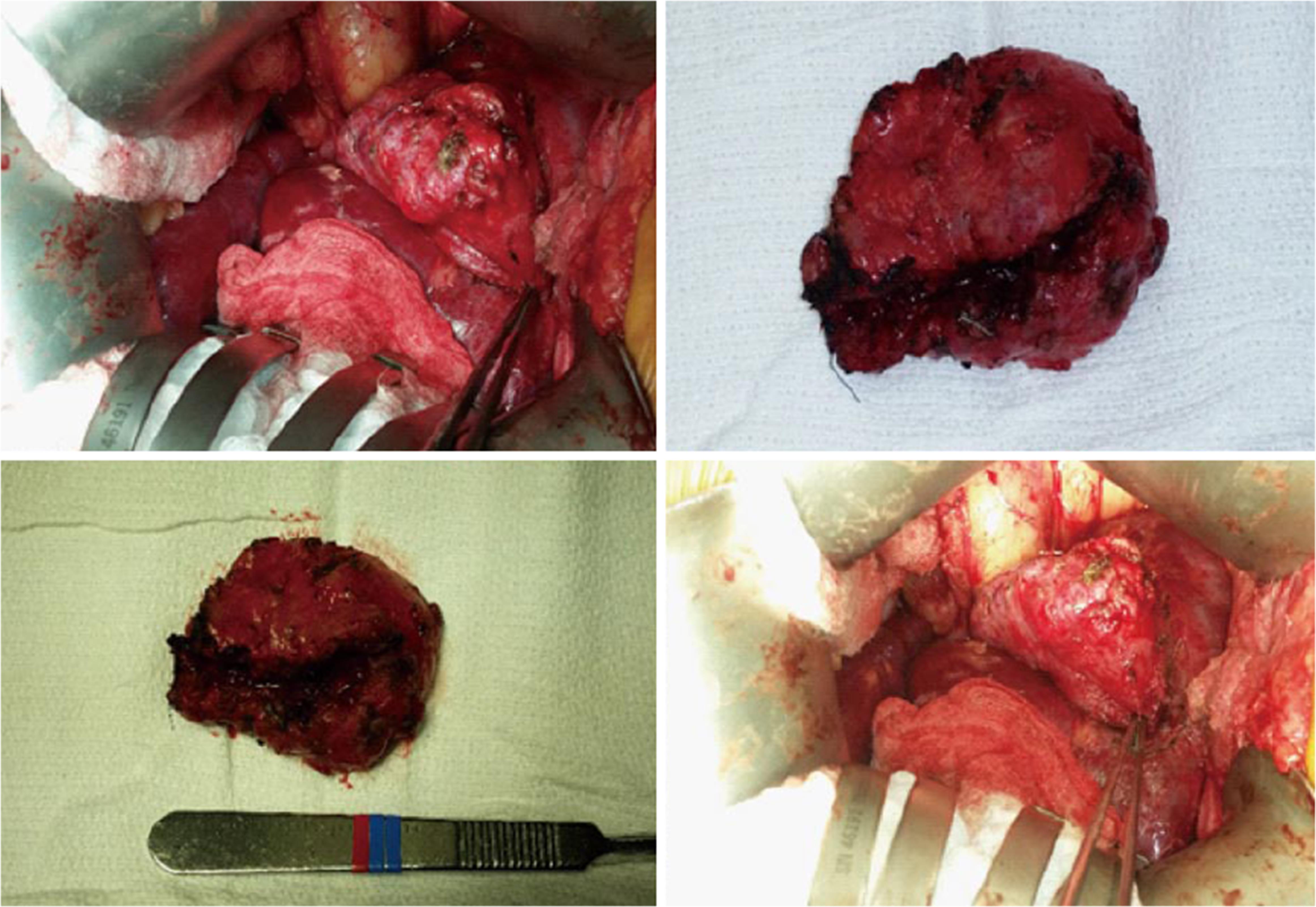
Figure 3 Intraoperative views and gross images of cystic duct mucocele.
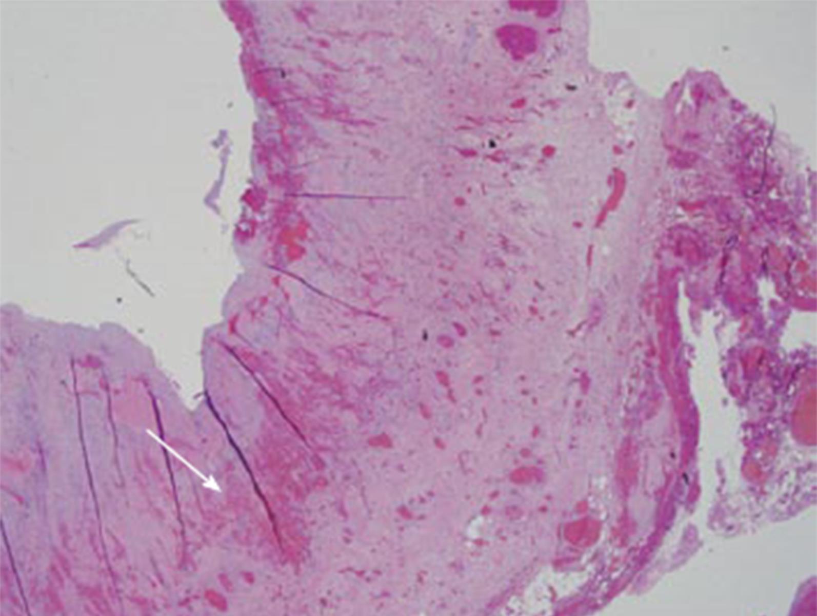
Figure 4 Section shows chronic cholangitis with prominent fibrosis, granulation tissue formation through mucosa, muscularis, and adventitia.
- Citation: Chaly T, Campsen J, O’Hara R, Hardman R, Gallegos-Orozco JF, Thiesset H, Kim RD. Mucocele mimicking a gallbladder in a transplanted liver: A case report and review of the literature. World J Transplant 2017; 7(6): 359-363
- URL: https://www.wjgnet.com/2220-3230/full/v7/i6/359.htm
- DOI: https://dx.doi.org/10.5500/wjt.v7.i6.359


