Copyright
©The Author(s) 2016.
World J Transplant. Sep 24, 2016; 6(3): 472-504
Published online Sep 24, 2016. doi: 10.5500/wjt.v6.i3.472
Published online Sep 24, 2016. doi: 10.5500/wjt.v6.i3.472
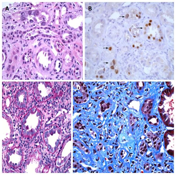
Figure 1 BK nephropathy in the native kidneys of a lung transplant recipient (Patient 2 in this report, A and B) and in the native kidneys of a bone marrow recipient (patient 1 in this report, C and D).
Kidney biopsy showing BK nephropathy (BKN), taken from a 70-year-old male with a history of lung transplantation for pulmonary fibrosis. A renal biopsy was performed because of significant worsening in renal function over a one-month period. A: Kidney biopsy showing active BK virus infection of renal tubules, with multiple homogeneous-appearing viral nuclear inclusions (white arrows), and features of associated acute tubular injury, including sloughing of tubular cells (H and E stain, 400 × magnification); B: Multiple renal tubules show positive nuclear staining for the SV40 large T antigen by immunoperoxidase staining (black arrows), confirming infection of tubular cells by polyomavirus (400 × magnification); Kidney biopsy from a 30-year-old male with a history of an allogeneic bone-marrow transplantation for aplastic anemia, who developed sequentially post-transplant Epstein-Barr virus associated large B-cell lymphoma, graft vs host disease and progressive elevation of his serum creatinine. This patient died from pneumococcal pneumonia and invasive aspergillosis two months after the diagnosis of BKN; C: Biopsy of renal cortex showing mononuclear tubulitis (black arrow), intranuclear BK virus inclusions (white arrow), and a prominent interstitial chronic inflammatory infiltrate (PAS stain, 400 × magnification); D: Another area of the biopsy shows extensive interstitial fibrosis and tubular atrophy, consistent with late changes secondary to infection (Trichrome stain, 400 × magnification).
- Citation: Vigil D, Konstantinov NK, Barry M, Harford AM, Servilla KS, Kim YH, Sun Y, Ganta K, Tzamaloukas AH. BK nephropathy in the native kidneys of patients with organ transplants: Clinical spectrum of BK infection. World J Transplant 2016; 6(3): 472-504
- URL: https://www.wjgnet.com/2220-3230/full/v6/i3/472.htm
- DOI: https://dx.doi.org/10.5500/wjt.v6.i3.472


