Copyright
©The Author(s) 2016.
World J Transplant. Jun 24, 2016; 6(2): 447-450
Published online Jun 24, 2016. doi: 10.5500/wjt.v6.i2.447
Published online Jun 24, 2016. doi: 10.5500/wjt.v6.i2.447
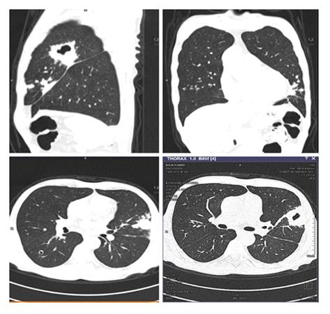
Figure 1 Multiple diffuse bilateral centrilobular and peribronchovascular cavitating nodules coalescing to form areas of consolidation with larger cavity in apico posterior segment of upper lobe of left lung.
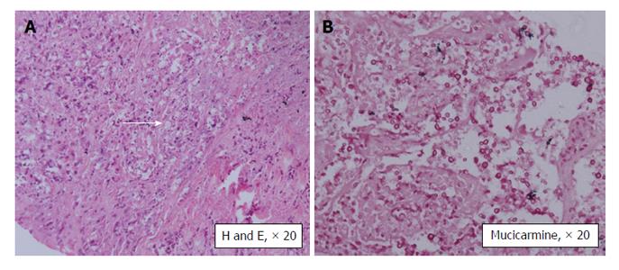
Figure 2 The histopathology showed cryptococcal infection.
Histopathology of the lung lesion shows: A: Large area of necrosis with numerous capsulated yeast forms of fungi (arrow) morphologically resembling Cryptococcus; B: Special histochemical stain (Mucicarmine) highlights its polysaccharide capsule.
- Citation: Subbiah AK, Arava S, Bagchi S, Madan K, Das CJ, Agarwal SK. Cavitary lung lesion 6 years after renal transplantation. World J Transplant 2016; 6(2): 447-450
- URL: https://www.wjgnet.com/2220-3230/full/v6/i2/447.htm
- DOI: https://dx.doi.org/10.5500/wjt.v6.i2.447


