Copyright
©The Author(s) 2016.
World J Transplant. Jun 24, 2016; 6(2): 278-290
Published online Jun 24, 2016. doi: 10.5500/wjt.v6.i2.278
Published online Jun 24, 2016. doi: 10.5500/wjt.v6.i2.278
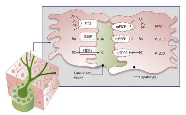
Figure 1 Disruption of bile flow and progressive familial intrahepatic cholestasis.
AP: Aminophospholipids; PS: Phosphatidylserine; PE: Phosphatidylethinolamine; BA: Bile acids; PC: Phosphatidylcholine; FIC1: Familial intrahepatic cholestasis protein 1; BSEP: Bile salt exporter pump; MDR3: Multidrug resistance protein 3; mFIC1: Mutant familial intrahepatic cholestasis protein 1; mBSEP: Mutant bile salt exporter pump; mMDR3: Mutant multidrug resistance protein; PFIC: Progressive familial intrahepatic cholestasis.
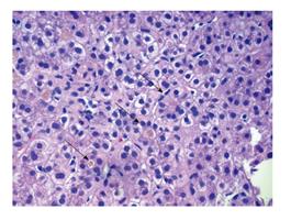
Figure 2 Progressive familial intrahepatic cholestasis type 1 with severe bland lobular cholestasis and lobular disarray.
The image shows bile plugging with surrounding pseudorosette formation (arrows). In PFIC1, the canalicular bile is course on electronic microscopy and also referred to as “Byler bile”. Thick bile is seen within the pseudorosette here on H and E stain. There is an absence of lobular inflammation and typically no features of neonatal giant cell hepatitis. PFIC: Progressive familial intrahepatic cholestasis.
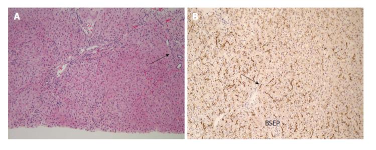
Figure 3 Progressive familial intrahepatic cholestasis type 2 is characterized by mutations in the ABCB11 gene.
A: Patients with progressive familial intrahepatic cholestasis type 2 (PFIC2) can initially present clinically similarly to PFIC1, but with more rapid progression of liver disease. Early on in the disease patients may present with neonatal giant cell hepatitis and lobular inflammation. However, there can be rapid progression with prominent duct reaction and progression to cirrhosis. This figure demonstrates prominent duct reaction in a patient with PFIC2 and advancing fibrosis (arrow). Duct reaction and cholestasis can also occur in patients with extrahepatic biliary obstruction so correlation with clinical findings is required; B: PFIC2 is also called BSEP disease and is characterized by mutations in the ABCB11 gene. ABCB11 encodes for the major canalicular bile salt exporter BSEP. Patients with normal BSEP expression show positive immunohistochemistry for BSEP with a canalicular pattern of staining (arrow). In some cases of PFIC2, there is complete lack of staining for BSEP. BSEP: Bile salt exporter pump.
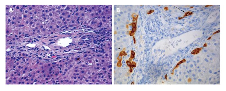
Figure 4 Progressive familial intrahepatic cholestasis type 1 and 2 can also present with duct paucity.
A and B: The portal tracts show an absence of bile duct with periportal duct reaction; B: A higher power view of the portal tract with vein on the left artery on the right (arrow) and no appreciable bile duct. Keratin 7 is negative in this portal tract in B and positive in the bile duct reaction (arrow) with some bile duct progenitor cells (paler brown staining arrowhead).
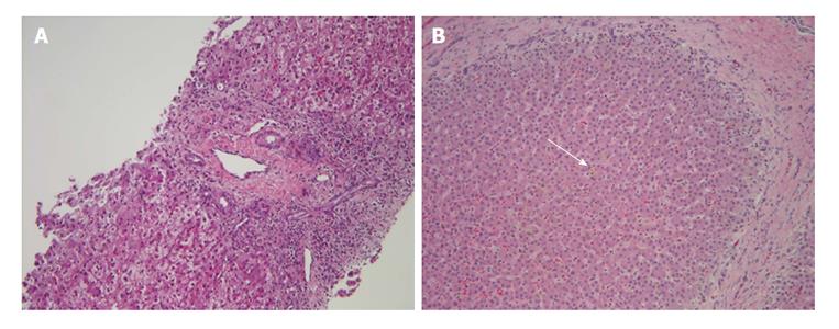
Figure 5 Clinical presentation of progressive familial intrahepatic cholestasis type 3.
A: Progressive familial intrahepatic cholestasis type 3 (PFIC3) has a variable clinical presentation and may show nonspecific biliary pattern of injury that can mimic extrahepatic biliary atresia such as bile duct proliferation and cholestasis. In this patient with PFIC3 there is cholestasis, inflammation, and bile duct proliferation; B: Biliary type cirrhosis in a patient with PFIC3 with severe cholestasis (arrow) and micronodular cirrhosis.
- Citation: Mehl A, Bohorquez H, Serrano MS, Galliano G, Reichman TW. Liver transplantation and the management of progressive familial intrahepatic cholestasis in children. World J Transplant 2016; 6(2): 278-290
- URL: https://www.wjgnet.com/2220-3230/full/v6/i2/278.htm
- DOI: https://dx.doi.org/10.5500/wjt.v6.i2.278


