Copyright
©The Author(s) 2016.
World J Transplant. Jun 24, 2016; 6(2): 272-277
Published online Jun 24, 2016. doi: 10.5500/wjt.v6.i2.272
Published online Jun 24, 2016. doi: 10.5500/wjt.v6.i2.272
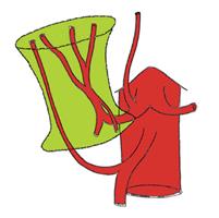
Figure 1 All the potential arterial branches that should be preserved during the recipient hepatectomy.
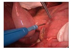
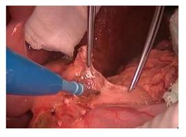
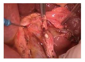
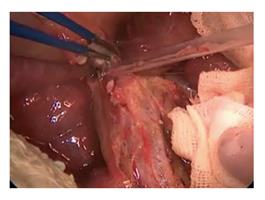
Figure 2 Opening small windows on the peritoneum of the distal hepatoduodenal ligament.
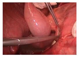
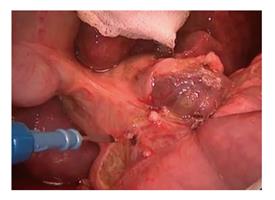
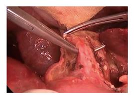
Figure 3 Dissection started just close to the duodenum.
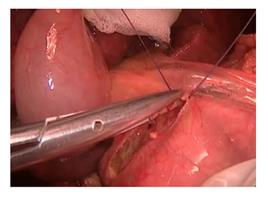
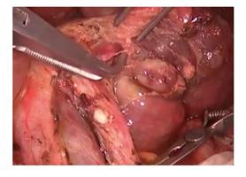
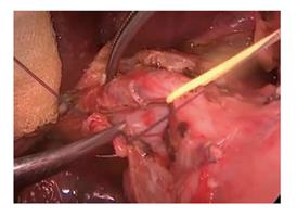
Figure 4 Vessels under the peritoneum were transected after ligation.
Avoiding electrocautery at this stage protects the duodenum from thermal injury.
Figure 5 The lymph node located at the infero-medial part of the hepatoduodenal ligament is an important landmark.
It locates along the upper border of the pancreas.
Figure 6 The landmark lymph node is one of the largest lymph nodes of the hepatoduodenal ligament.
Its Identification enables to find out the arteria hepatica communis which is just under this lymph node. The dissection is extended to the infero-lateral part of the hepatoduodenal ligament by jumping over the distal common bile duct. Another large counterpart lymph node is exposed near to the distal common bile duct.
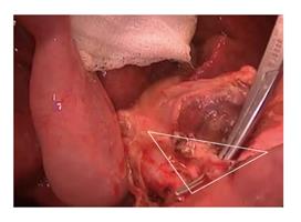
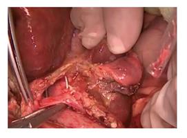
Figure 7 The gastro-duodenal artery is identified in the triangle composed of; distal common bile duct, hepatic artery and upper border of the duodenum.
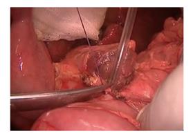
Figure 8 Ligation and transaction of the gastroduodenal artery.
Figure 9 Division of the gastroduodenal artery makes the portal vein visible.
Figure 10 Bulldog clamp applied to the proximal common hepatic artery to prevent the intimal damage in the artery during further dissections.
Figure 11 Right hepatic artery is crossing the common bile duct from the posterior.
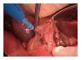
Figure 12 Traction of the cystic duct stump ensures a safer dissection of the lateral border of the common bile duct.
Figure 13 Removing the sheet over the extrahepatic bile ducts provides the identification of the medial and lateral borders of the bile ducts.
Figure 14 Common bile duct is further liberated and hanged by the right angle clamp.
Figure 15 Medial traction applied to the bile duct by a rubber band and the lymphatics are transected.
This transaction should be done after being sure that there is no accessory right hepatic artery arising from the superior mesenteric artery.
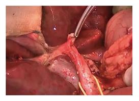
Figure 16 Removing the lymph nodes and the transection of the arteries enables a well visualized and prolonged common bile duct and portal vein.
- Citation: Kayaalp C, Tolan K, Yilmaz S. Hepatoduodenal ligament dissection technique during recipient hepatectomy for liver transplantation: How I do it? World J Transplant 2016; 6(2): 272-277
- URL: https://www.wjgnet.com/2220-3230/full/v6/i2/272.htm
- DOI: https://dx.doi.org/10.5500/wjt.v6.i2.272


