Copyright
©The Author(s) 2016.
World J Transplant. Mar 24, 2016; 6(1): 249-254
Published online Mar 24, 2016. doi: 10.5500/wjt.v6.i1.249
Published online Mar 24, 2016. doi: 10.5500/wjt.v6.i1.249
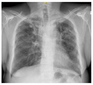
Figure 1 Chest X-ray postro-anterior view at 15 years.
Note right upper zone nodular and interstitial opacities.
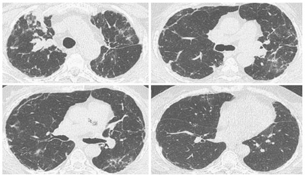
Figure 2 Computed tomography of the chest.
RUL nodules with bilateral interstitial thickening and scattered ground glass opacities.
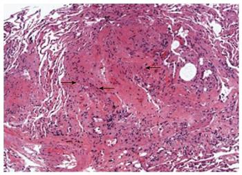
Figure 3 Histopathological examination of the transbronchial biopsy revealing spindle shaped lymphangioleiomyomatosis (arrows) cells suggestive of recurrence.
- Citation: Zaki KS, Aryan Z, Mehta AC, Akindipe O, Budev M. Recurrence of lymphangioleiomyomatosis: Nine years after a bilateral lung transplantation. World J Transplant 2016; 6(1): 249-254
- URL: https://www.wjgnet.com/2220-3230/full/v6/i1/249.htm
- DOI: https://dx.doi.org/10.5500/wjt.v6.i1.249


