Copyright
©The Author(s) 2016.
World J Transplant. Mar 24, 2016; 6(1): 10-27
Published online Mar 24, 2016. doi: 10.5500/wjt.v6.i1.10
Published online Mar 24, 2016. doi: 10.5500/wjt.v6.i1.10
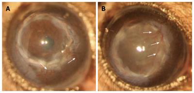
Figure 1 Corneal allografts in C57BL/6 mice.
(A) Accepted and (B) rejected corneal allografts (Balb/c donor) in C57BL/6 mice demonstrating invasion of blood vessels (arrows); the rejected graft shows more blood vessels invading the donor graft.
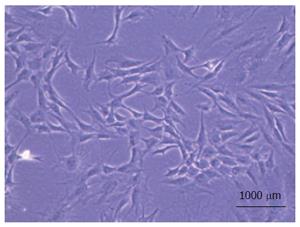
Figure 2 Spindle shaped morphology characteristic of multipotent mesenchymal stem cells.
Figure shows passage 4 mesenchymal stem cells derived from the non-haematopoietic sub-population of bone marrow harvested from 6-8 wk old Balb/c mice.
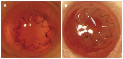
Figure 3 Clinical images of tissue engineered collagen-based hydrogels transplanted by full-thickness keratoplasty into naïve Balb/c mice at different time points post grafting.
A: Clear hydrogel 1 d post transplantation; B: Hydrogel clarity is reduced 9 d post transplantation due to retro-hydrogel membrane formation (from periphery towards central cornea as indicated by arrows).
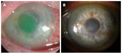
Figure 4 Clinical images of a “high-risk” cornea from a patient with ocular surface disease transplanted with a recombinant human collagen-based hydrogel.
A: Corneal graft bed showing fluorescein stained epithelial erosion/ulcer (green/yellow staining) and vascularization before transplantation; B: Relatively clear cornea with clinically visible regression of peripheral corneal vessels 12 mo post-surgery[215].
- Citation: Yu T, Rajendran V, Griffith M, Forrester JV, Kuffová L. High-risk corneal allografts: A therapeutic challenge. World J Transplant 2016; 6(1): 10-27
- URL: https://www.wjgnet.com/2220-3230/full/v6/i1/10.htm
- DOI: https://dx.doi.org/10.5500/wjt.v6.i1.10


