Copyright
©The Author(s) 2015.
World J Transplant. Dec 24, 2015; 5(4): 360-365
Published online Dec 24, 2015. doi: 10.5500/wjt.v5.i4.360
Published online Dec 24, 2015. doi: 10.5500/wjt.v5.i4.360
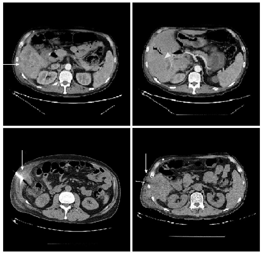
Figure 1 Computed tomography scan images of the liver.
The vertical white arrows show the site of needle biopsy. The horizontal white arrow shows tumour mass in S5 extending to S6 and the arrow head shows the site of right intrahepatic duct dilatation.
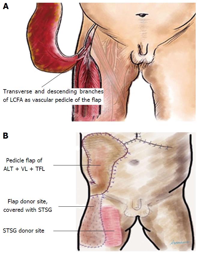
Figure 2 Diagramtic depiction of the myocutaneous pedicle flap for abdominal wall reconstruction.
A: Extended right thigh flap based on the transverse and descending branches of the LCFA; B: Pedicle flap of ALT + VL + TFL for coverage of right abdominal wall defect. Donor site covered with STSG taken from the right thigh. LCFA: Lateral circumflex femoral artery; ALT: Anterolateral thigh; VL: Vastus lateralis; TFL: Tensor fascia latae; STSG: Split thickness skin graft.
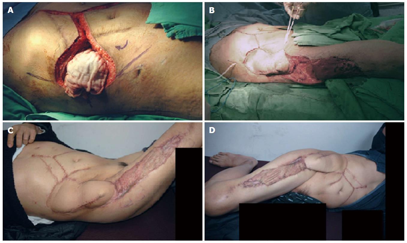
Figure 3 Recipient’s intraoperative and follow up images.
A: Ten centimeter × 10 cm diameter right abdominal wall defect following the wide local excision of the area; B: Perioperative picture of the transposition of the right thigh extended pedicle flap and coverage of the right side abdominal wall defect; C: Post operative picture from the outpatient clinic on a three months follow up; D: Post operative picture from the outpatient clinic on a six months follow up.
- Citation: Yang HR, Thorat A, Gesakis K, Li PC, Kiranantawat K, Chen HC, Jeng LB. Living donor liver transplantation with abdominal wall reconstruction for hepatocellular carcinoma with needle track seeding. World J Transplant 2015; 5(4): 360-365
- URL: https://www.wjgnet.com/2220-3230/full/v5/i4/360.htm
- DOI: https://dx.doi.org/10.5500/wjt.v5.i4.360


