Copyright
©The Author(s) 2015.
World J Transplant. Dec 24, 2015; 5(4): 354-359
Published online Dec 24, 2015. doi: 10.5500/wjt.v5.i4.354
Published online Dec 24, 2015. doi: 10.5500/wjt.v5.i4.354
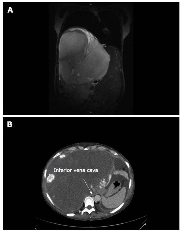
Figure 1 Radiological imaging.
A: Radiological imaging showing a tumour of 21.7 cm × 23.7 cm × 25.5 cm in size in segments V-VIII. The tumour volume was 6000 mL; the total liver volume was calculated as 9691 mL; B: The vena cava inferior was massively dislocated to the left and slit-shaped due to compression. This contributes to progressive ascites.
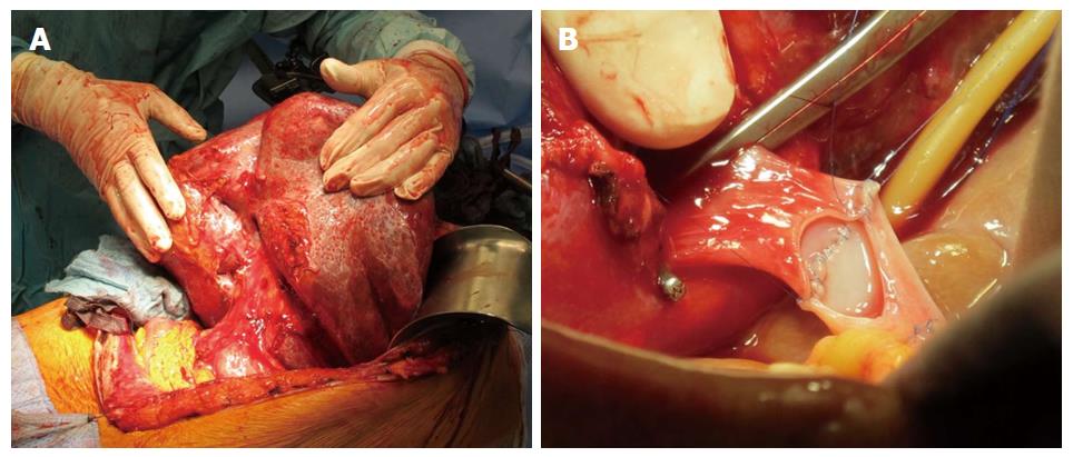
Figure 2 Recipient liver before explantation and portal vein anastomosis.
A: In February 2014, orthotopic liver transplantation was performed. The large size of the donor liver was tolerated because of the enlarged liver size of the patient; B: The main stem of the patient’s portal vein showed no thrombosis. Bicaval anastomosis followed by portal vein anastomosis was performed. The arterial anastomosis was performed to the recipient’s gastroduodenal artery. Biliary drainage was achieved by choledochocholedochostomy. Age of the donor: 36 years; cold ischemia time: 11 h and 54 min.
- Citation: Lange UG, Bucher JN, Schoenberg MB, Benzing C, Schmelzle M, Gradistanac T, Strocka S, Hau HM, Bartels M. Orthotopic liver transplantation for giant liver haemangioma: A case report. World J Transplant 2015; 5(4): 354-359
- URL: https://www.wjgnet.com/2220-3230/full/v5/i4/354.htm
- DOI: https://dx.doi.org/10.5500/wjt.v5.i4.354


