Copyright
©The Author(s) 2015.
World J Transplant. Dec 24, 2015; 5(4): 300-309
Published online Dec 24, 2015. doi: 10.5500/wjt.v5.i4.300
Published online Dec 24, 2015. doi: 10.5500/wjt.v5.i4.300
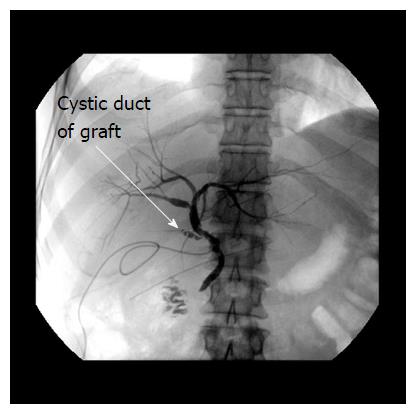
Figure 1 Normal anatomy of bile duct anastomosis (side-to-side): T-tube X-ray six weeks after liver transplantation.
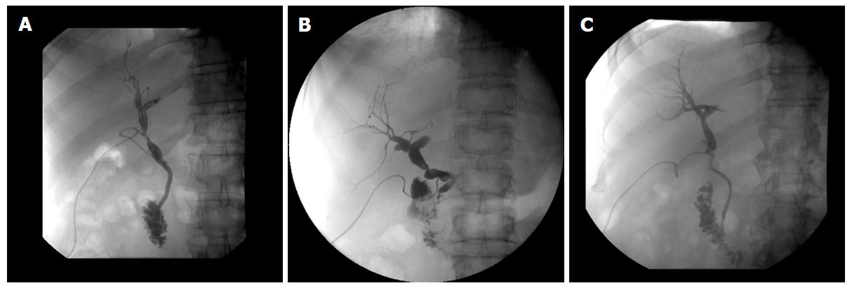
Figure 2 Different types of bile duct anastomotic pathologies: All T-tube X-rays six weeks after liver transplantation.
A: Stenosis (> 30%) after side-to-side anstomosis, resolved after endoscopic stent treatment for 3 mo; B: Stenosis (> 30%) after end-to-end anstomosis, all lab values normal, no clinical relevance, no intervention; C: No anastomotic stenosis but incongruence of graft- and recipient bile duct, no clinical relevance, normal lab values, surveillance.
- Citation: Kienlein S, Schoening W, Andert A, Kroy D, Neumann UP, Schmeding M. Biliary complications in liver transplantation: Impact of anastomotic technique and ischemic time on short- and long-term outcome. World J Transplant 2015; 5(4): 300-309
- URL: https://www.wjgnet.com/2220-3230/full/v5/i4/300.htm
- DOI: https://dx.doi.org/10.5500/wjt.v5.i4.300


