Copyright
©The Author(s) 2015.
World J Transplant. Dec 24, 2015; 5(4): 165-182
Published online Dec 24, 2015. doi: 10.5500/wjt.v5.i4.165
Published online Dec 24, 2015. doi: 10.5500/wjt.v5.i4.165
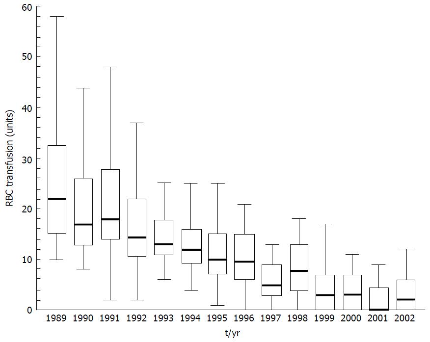
Figure 1 Red blood cell transfusion requirement in patients undergoing primary liver transplantation at the University Medical Centre, Groningen from 1989 to 2002.
Data presented as box and whisker plots representing median, interquartile range and 5%-95% range. Both variation in transfusion and median number of RBC transfused has declined over time. From Porte et al[6] 2004. RBC: Red blood cell.
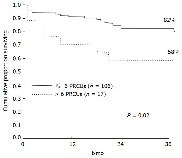
Figure 2 Kaplan meier survival curve demonstrating reduced survival in patients who received > 6 units red blood cell up to 36 mo post liver transplantation.
From Ramos et al[13]. PRCUs: Packed red cell units.
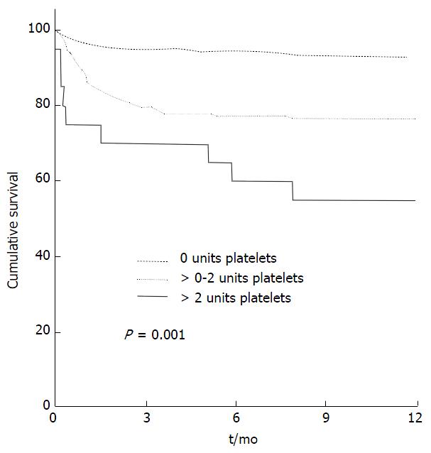
Figure 3 Kaplan Meier curve representing cumulative survival with transfusion of no, 0-2 units and > 2 units of platelets.
From de Boer et al[14] 2008.
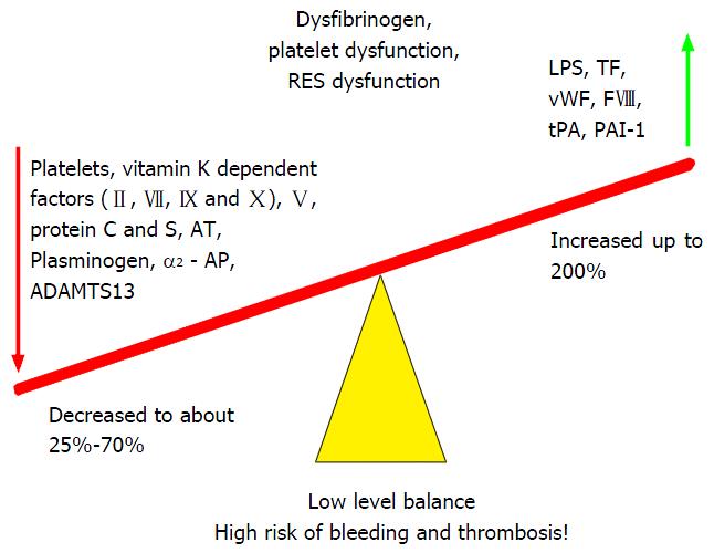
Figure 4 Fragile rebalancing of pro and anti-haemostatic factors with limited reserves leads to increased thrombotic and bleeding tendency.
From Saner et al[43] 2013. TF: Tissue factor; vWF: Von Willebrand Factor; FVIII: Factor VIII; tPA: Tissue plasminogen activator; PAI: Plasminogen activator inhibitor; ADAMTS13: A disintegrin and metalloproteinase with a thrombospondin type 1 motif, member 13; AT: Antithrombin; LPS: Lipopolysaccharide.
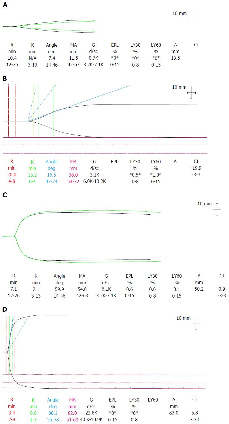
Figure 5 Thromboelastography traces depicting.
A: Dilutional coagulopathy with loss of endogenous thrombin generating potential, low fibrinogen and platelets seen as a typical “arrowhead” trace; B: Hypocoagulable state with prolonged R time but retention of thrombin generation; C: Normal coagulation profile; D: Hypercoagulability with a short R time, steep rate of clot formation (α angle) and increased clot MA. R: Reaction time; K: Kinetics time taken to achieve a certain level of clot strength (20 mm); MA: Maximum amplitude; G: Log derivation of the MA; EPL: Estimated percent lysis; Ly30: Lysis at 30 min; Ly60: Lysis at 60 min; CI: Coagulation index.
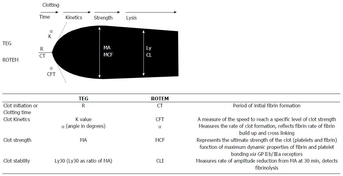
Figure 6 Schematic of thromboelastography/rotational thromboelastometry parameters.
TEG: Thromboelastography; K time: Coagulation time (20-mm clot); Ly30: Lysis at 30 min; MA: Maximum amplitude; R time: Reaction time (2-mm clot); ROTEM: Rotational thromboelastometry; CFT: Clot formation time (20-mm clot); CLI: Clot lysis index; CT: Clotting time (2-mm clot); MCF: Maximum clot firmness; ML: Maximal lysis. From Mallett[37] 2015.
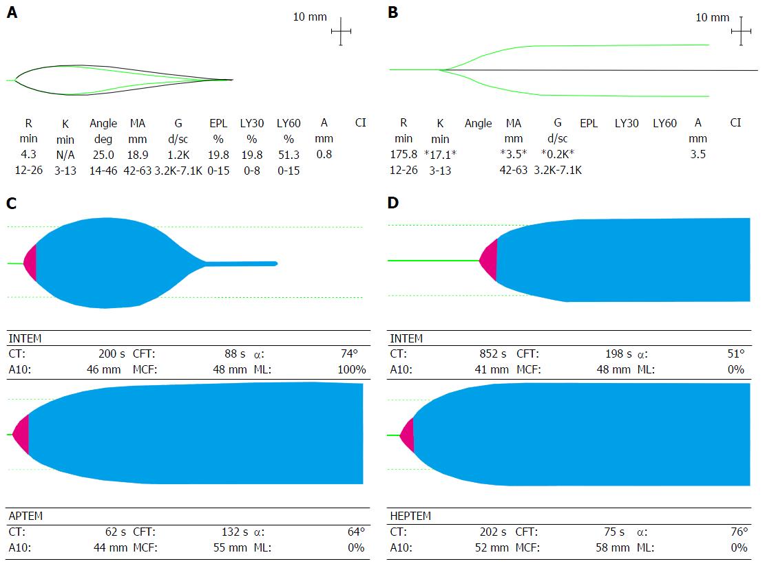
Figure 7 Viscoelastic traces depicting hyperfibrinolysis (A) on thromboelastography and (C) on rotational thromboelastometry with aprotinin thromboelastometry test (reversal of fibrinolysis with aprotinin); Viscoelastic traces depicting heparin-like effect (B) on thromboelastography and (D) on Rotational thromboelastometry with reversal of heparin-like effect on heparinase modified thromboelastometry.
APTEM: Aprotinin thromboelastometry; HEPTEM: Heparinase modified thromboelastometry; INTEM: Intrinsic thromboelastometry; CT: Clotting time; CFT: Clot formation time; A10: Clot firmness (amplitude) at 10 min; MCF: Maximum clot firmness; ML: Maximum lysis.
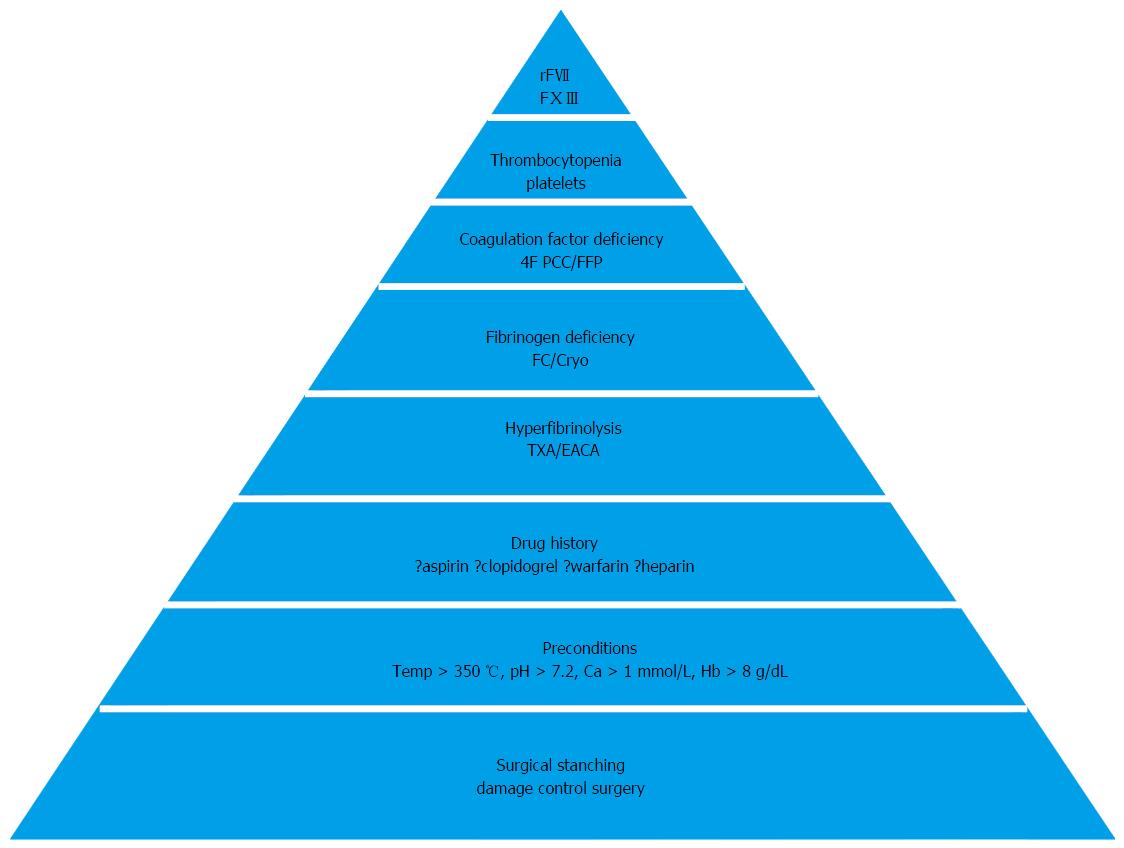
Figure 8 Pyramid of therapy in coagulopathy with sequence of hemostatic therapeutic interventions (from base to tip).
Ca: Calcium; Hb: Haemoglobin; TXA: Tranexamic acid; EACA: Epsilon aminocaproic acid; FC: Fibrinogen concentrate; Cryo: Cryoprecipitate; rFVII: Recombinant activated factor VII; FXIII: Factor XIII. From Görlinger et al[79] 2006.
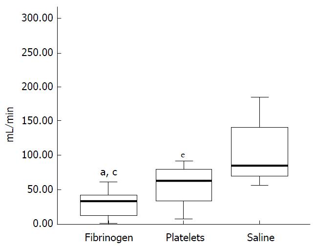
Figure 9 Rate of blood loss (mL/min) in thrombocytopenic pigs with an induced haemorrhagic liver injury infused with fibrinogen concentrate, platelet concentrate or saline.
Lowest rate of blood loss seen in pigs infused with fibrinogen concentrate. aP < 0.05 fibrinogen vs platelet group, cP < 0.05 fibrinogen vs saline group, eP < 0.05 platelet vs saline group. From Velik-Salchner et al[103] 2007.
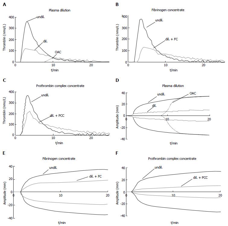
Figure 10 Effect of plasma dilution and factor deficiency on thrombin generation and fibrin clot formation.
Representative curves of tissue factor - induced thrombin generation (A-C) and fibrin clot formation (D-F) of undiluted normal plasma (undil.), 5 × diluted plasma (dil.), and plasma from patient taking oral anticoagulants (OAC, INR = 4). Effects of supplementing diluted plasma with fibrinogen concentrate (+ FC): Increase in clot strength with no effect on thrombin generation (B, E). Effects of supplementing diluted plasma with prothrombin complex concentrate (+ PCC): Increase in thrombin generation but no effect on clot strength (C, F). From Schols[105] 2010. OAC: Oral anticoagulatnts; INR: International normalized ratio.
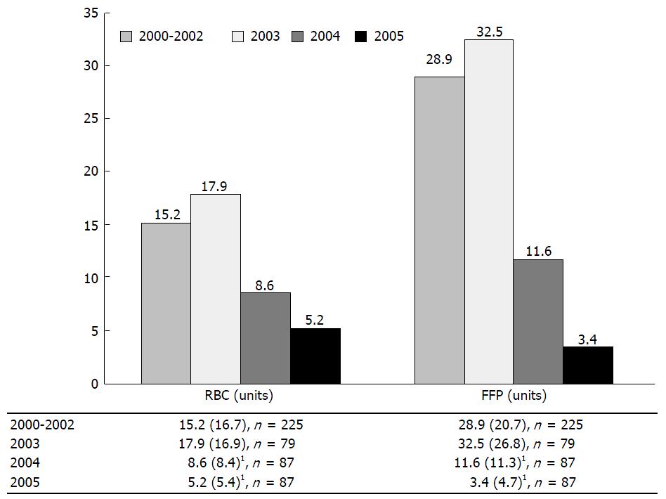
Figure 11 Blood transfusion during liver transplantation per case: Bar plots of red blood cells and fresh frozen plasma by year.
1Indicates a significant difference from the 2000-2002 value at a significance level of P = 0.05 based on the Wald t-test for the respective regression coefficient with adjustments for age, gender, baseline weight and height. FFP: Fresh frozen plasma; n: Samples size; RBC: Red blood cells. From Hevesi et al[125] 2009.
- Citation: Donohue CI, Mallett SV. Reducing transfusion requirements in liver transplantation. World J Transplant 2015; 5(4): 165-182
- URL: https://www.wjgnet.com/2220-3230/full/v5/i4/165.htm
- DOI: https://dx.doi.org/10.5500/wjt.v5.i4.165


