Copyright
©2014 Baishideng Publishing Group Inc.
World J Transplant. Jun 24, 2014; 4(2): 148-152
Published online Jun 24, 2014. doi: 10.5500/wjt.v4.i2.148
Published online Jun 24, 2014. doi: 10.5500/wjt.v4.i2.148
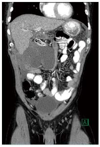
Figure 1 Desmoid tumor at initial presentation.
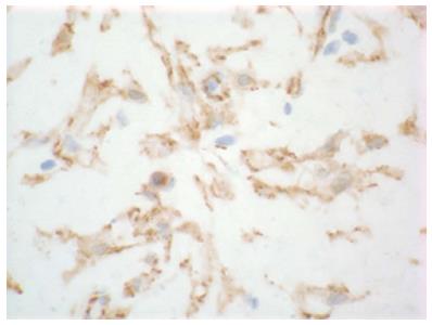
Figure 2 Immunostained slide of tumor histology, demonstrating low cellularity tumor in a myxoid matrix with positive beta-catenin staining.
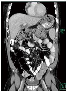
Figure 3 Imaging 18 mo after excision.
- Citation: Fleetwood VA, Zielsdorf S, Eswaran S, Jakate S, Chan EY. Intra-abdominal desmoid tumor after liver transplantation: A case report. World J Transplant 2014; 4(2): 148-152
- URL: https://www.wjgnet.com/2220-3230/full/v4/i2/148.htm
- DOI: https://dx.doi.org/10.5500/wjt.v4.i2.148


