Copyright
©2014 Baishideng Publishing Group Inc.
World J Transplant. Jun 24, 2014; 4(2): 111-121
Published online Jun 24, 2014. doi: 10.5500/wjt.v4.i2.111
Published online Jun 24, 2014. doi: 10.5500/wjt.v4.i2.111
Figure 1 Line diagram showing layers of cryopreserved human amniotic membrane, oriented with the stromal side in contact with the nitrocellulose filter paper and epithelial side facing up.
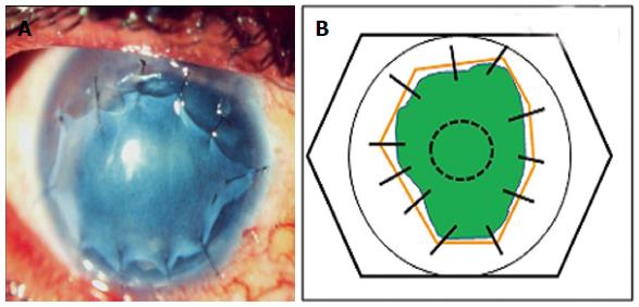
Figure 2 (A) Amniotic membrane used as an inlay graft (B) line diagram showing amniotic membrane (solid orange lines) used as an Inlay graft for a non-healing epithelial defect (green).
The membrane is trimmed to fit the size of the underlying defect and sutured to the cornea using interrupted 10-0 nylon monofilament sutures.
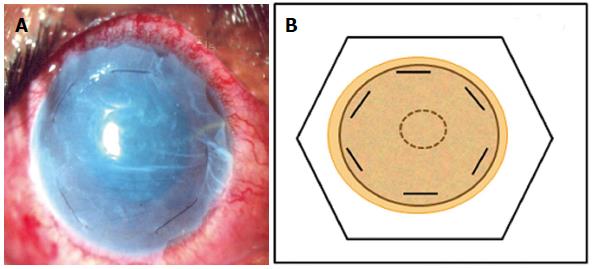
Figure 3 Amniotic membrane used as an overlay patch.
A: Clinical photograph; B: Line diagram. The amniotic membrane is trimmed to cover the whole of the corneal surface and fixed by 10-0 nylon monofilamemt sutures at the corneal periphery parallel to the limbus.
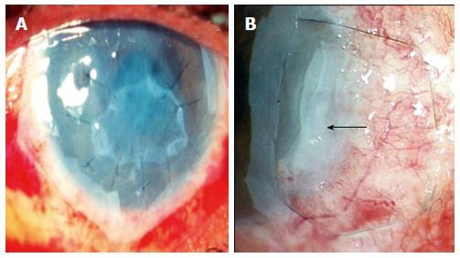
Figure 4 Single layer inlay covered by a larger “patch” or “onlay” (A), multi-layer inlay ( amniotic membrane folded upon itself in form of a blanket fold- black arrow) covered by a larger “patch” which is fixed to underlying episclera by a purse string suture in a case of deep, non-resolving peripheral ulcerative keratitis (B).
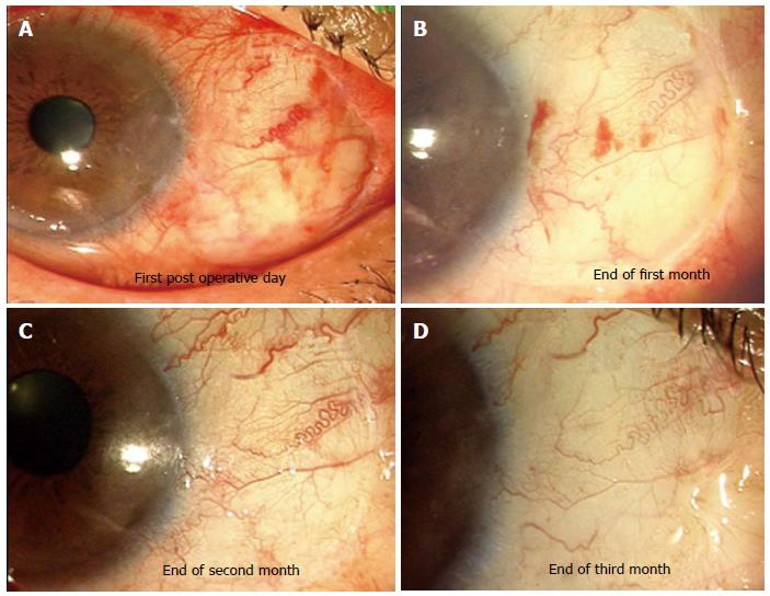
Figure 5 Amniotic membrane used to cover bare sclera after excision of primary pterygium.
A-D: Serial photographs showing appearance of amniotic membrane graft. At end of 3 mo excellent integration and cosmetic appearance was achieved.
- Citation: Malhotra C, Jain AK. Human amniotic membrane transplantation: Different modalities of its use in ophthalmology. World J Transplant 2014; 4(2): 111-121
- URL: https://www.wjgnet.com/2220-3230/full/v4/i2/111.htm
- DOI: https://dx.doi.org/10.5500/wjt.v4.i2.111


