Copyright
©The Author(s) 2025.
World J Transplant. Mar 18, 2025; 15(1): 97598
Published online Mar 18, 2025. doi: 10.5500/wjt.v15.i1.97598
Published online Mar 18, 2025. doi: 10.5500/wjt.v15.i1.97598
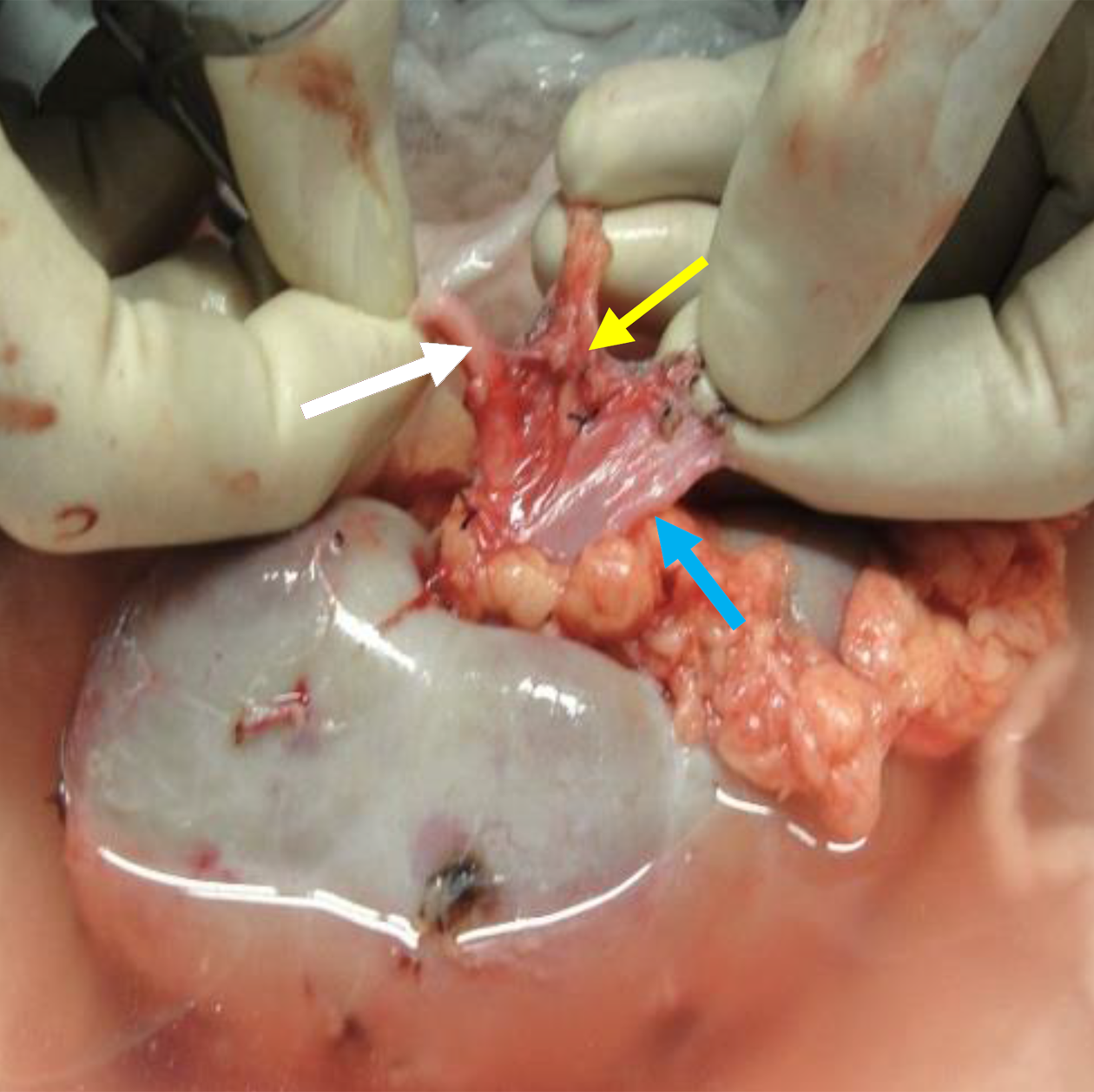
Figure 1 Right renal vein circumferential mobilization.
Lymphatic tissue (yellow arrow) between the artery (white arrow) and the vein (blue arrow), needs to be excised to mobilize the vein.
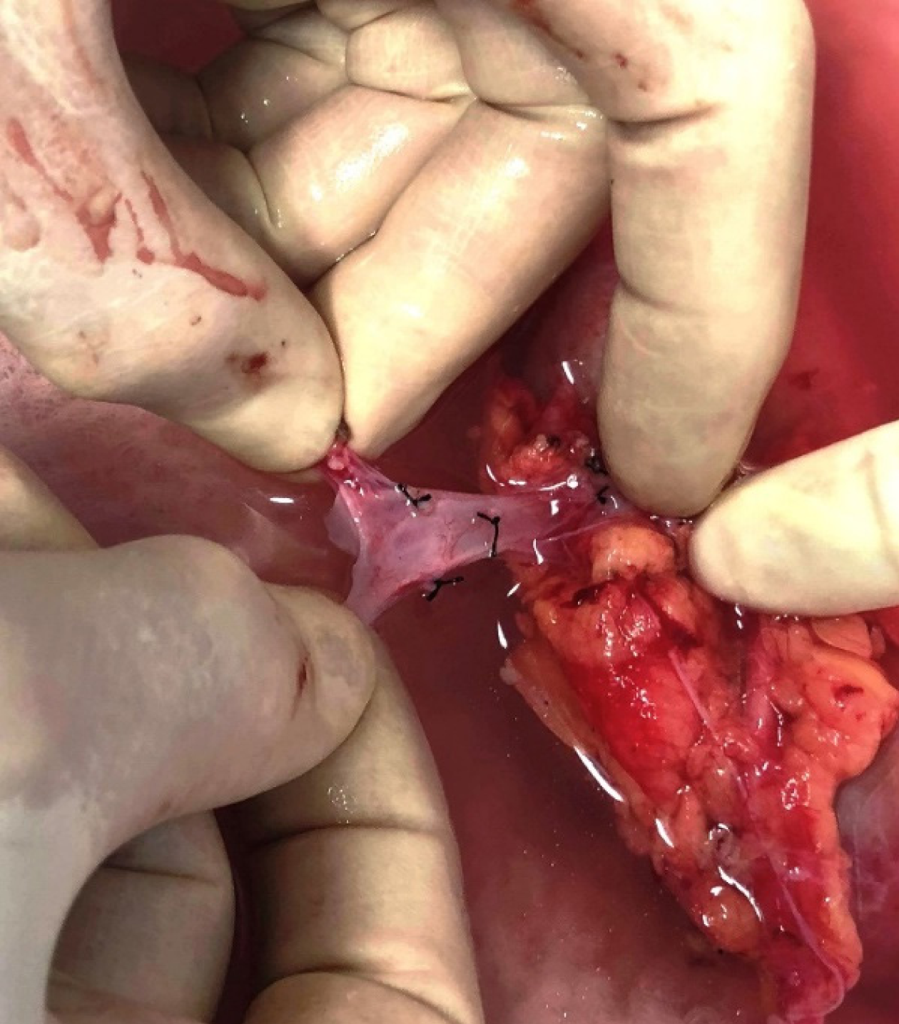
Figure 2 Thin-walled, fragile right renal vein on full stretch.
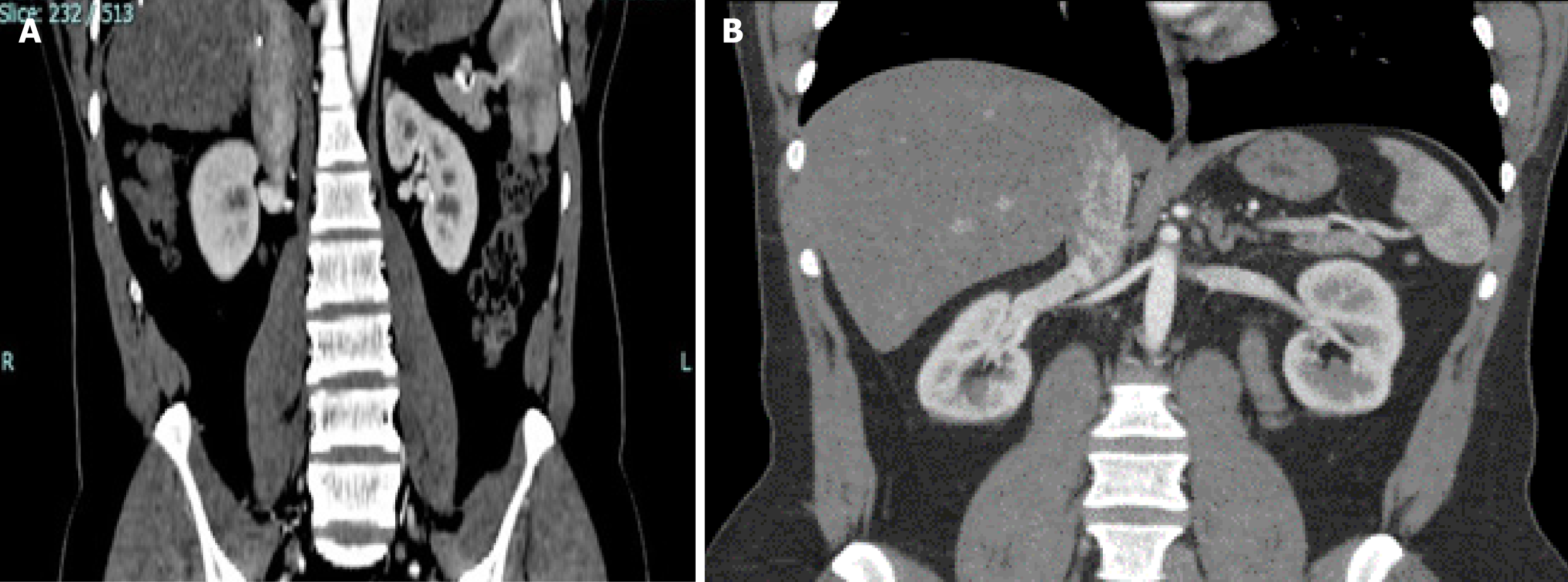
Figure 3 Computed tomography images showing right renal vein entry into the inferior vena cava.
A: Short vein in horizontal entry because perpendicular is the shortest distance; B: Long vein in obtuse angle entry.
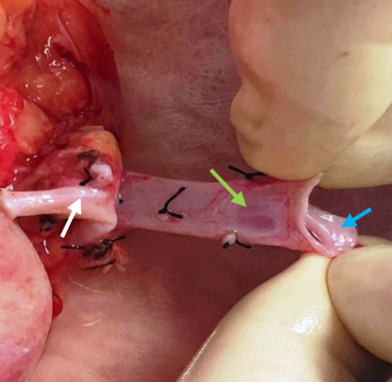
Figure 4 Lengthened right renal vein.
The weak oval area is clearly visible (green arrow) and silk ligatures on tissue around the vein and artery (white arrow) are the result of circumferential dissection to mobilize the vein. The caval cuff (blue arrow) was used for suturing to avoid weak area.
- Citation: Khan T, Ahmad N, Iqbal Q, Hassan M, Asnath L, Khan N, Shakeel S. Comparative study of living donor kidney transplants: Right vs left. World J Transplant 2025; 15(1): 97598
- URL: https://www.wjgnet.com/2220-3230/full/v15/i1/97598.htm
- DOI: https://dx.doi.org/10.5500/wjt.v15.i1.97598


