Copyright
©The Author(s) 2024.
World J Transplant. Mar 18, 2024; 14(1): 88938
Published online Mar 18, 2024. doi: 10.5500/wjt.v14.i1.88938
Published online Mar 18, 2024. doi: 10.5500/wjt.v14.i1.88938
Figure 1 Doppler ultrasound evaluation of the hepatic artery.
A: Intercostal color and spectral doppler image of a normal hepatic artery at the porta hepatis in the liver graft of a 51-year-old woman on postoperative day 3 after transplant, depicting a rapid systolic upstroke with continuous low-velocity diastolic flow and a normal resistive index; B: Subcostal color doppler image of the right hepatic lobe in a 46-year-old man on postoperative day 2 demonstrates vascular flow in the portal vein, with no hepatic artery flow detected on color or spectral doppler images at the porta hepatis.
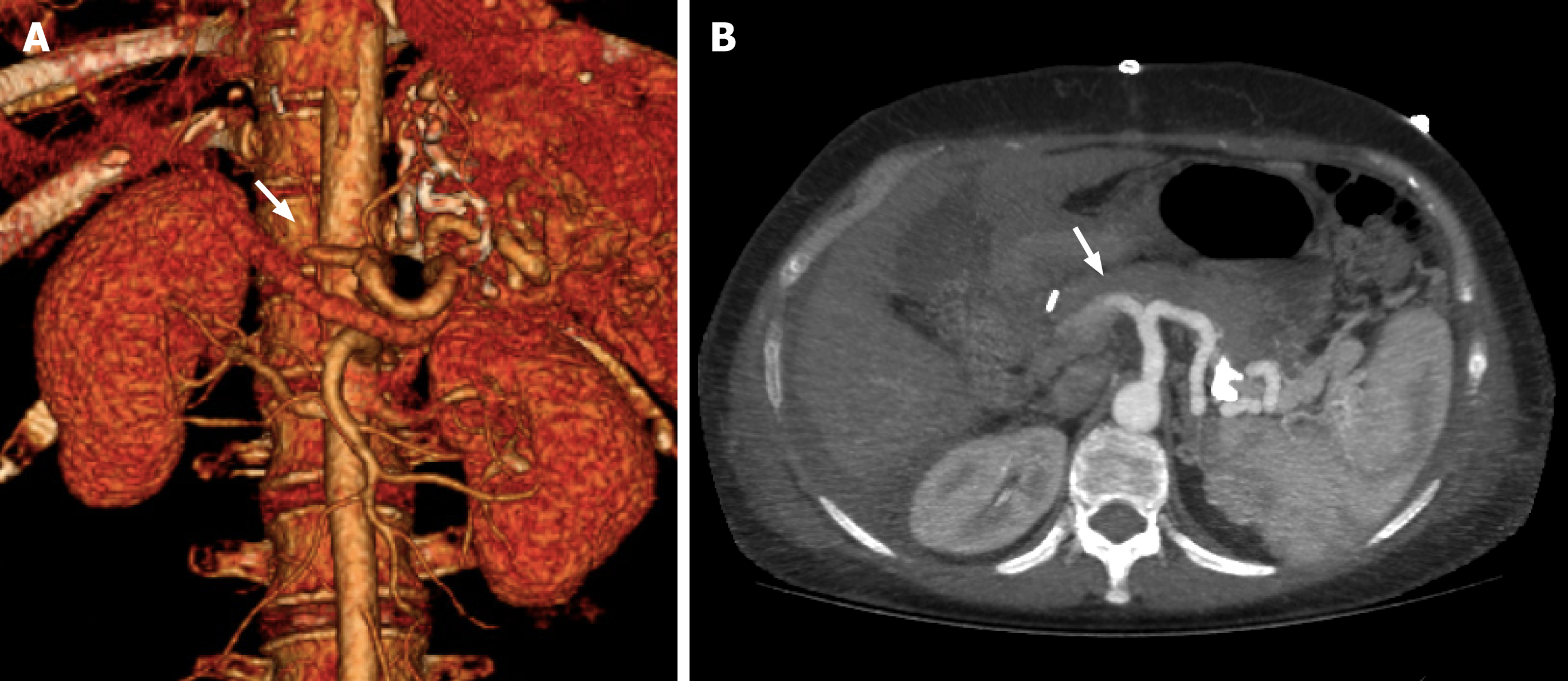
Figure 2 Computed tomography angiography evaluation of hepatic artery thrombosis in liver graft.
A 51-year-old woman on postoperative day 7 after a liver transplant. A: Axial abdominal computed tomography angiography (CTA) images at maximum intensity projection (white arrow); B: Coronal 3D volume rendering CTA reconstruction showing absence of vascular opacification of vessels distal to occlusion of the hepatic artery (white arrow).
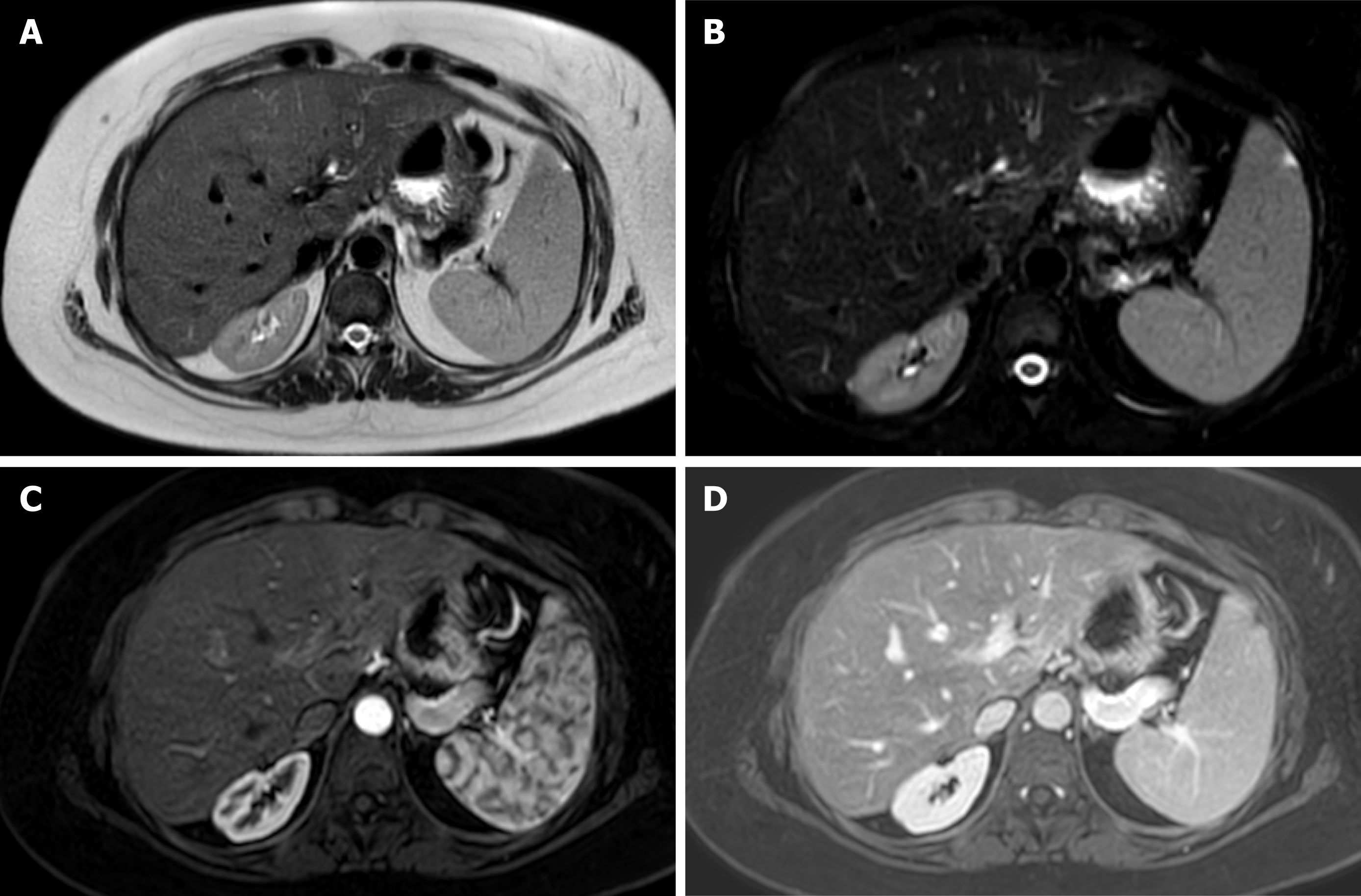
Figure 3 Magnetic resonance imaging of liver graft.
Axial T2-weighted single-shot fast spin-echo image. A: Fat saturation (Fat-sat); B: Contrast enhanced T1-weighted gradient-echo image in late arterial; C: Portal phase; D: Depicts a homogeneous signal intensity at graft parenchyma, with adequate representation of intrahepatic and extrahepatic arterial branches, venous vessels and biliary ducts, in a 32-year-old man on postoperative surveillance after a liver transplantation.
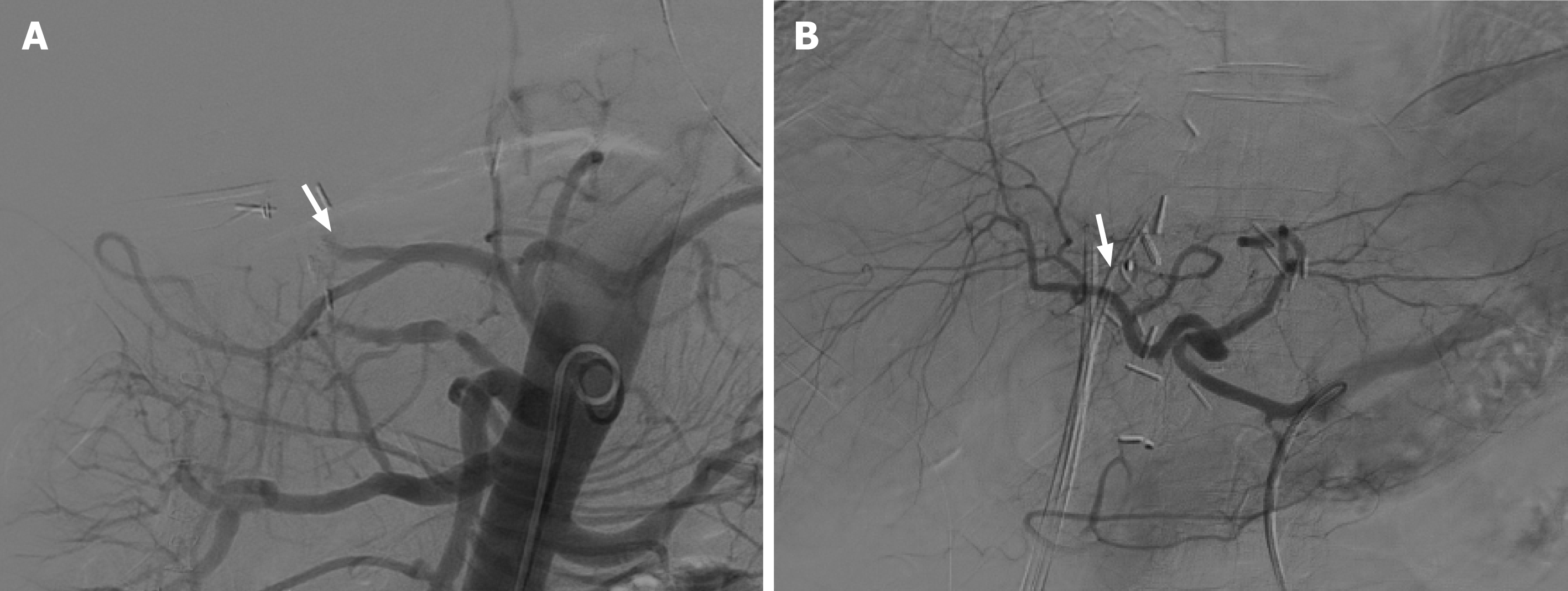
Figure 4 Visceral angiography performed 3 d after orthotopic liver transplant.
A: Demonstrated complete occlusion of the hepatic artery (white arrow); B: Recanalization of the hepatic artery after thrombectomy, with improved intrahepatic blood flow (white arrow).
- Citation: Lindner C, Riquelme R, San Martín R, Quezada F, Valenzuela J, Maureira JP, Einersen M. Improving the radiological diagnosis of hepatic artery thrombosis after liver transplantation: Current approaches and future challenges. World J Transplant 2024; 14(1): 88938
- URL: https://www.wjgnet.com/2220-3230/full/v14/i1/88938.htm
- DOI: https://dx.doi.org/10.5500/wjt.v14.i1.88938


