Copyright
©The Author(s) 2022.
World J Transplant. Jul 18, 2022; 12(7): 163-174
Published online Jul 18, 2022. doi: 10.5500/wjt.v12.i7.163
Published online Jul 18, 2022. doi: 10.5500/wjt.v12.i7.163
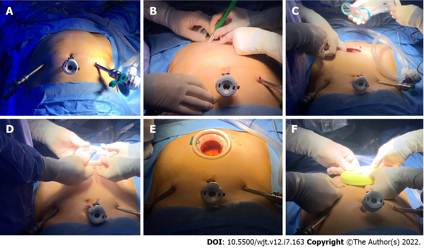
Figure 1 Overview of the main steps for Alexis® Wound Protectors/Retractors placement through Pfannestiel incision according to the University of Florence technique for robot-assisted kidney transplantation.
A: After ports placement; B and C: A Pfannestiel incision is performed; D-F: The Alexis® device is placed through Pfannestiel incision.
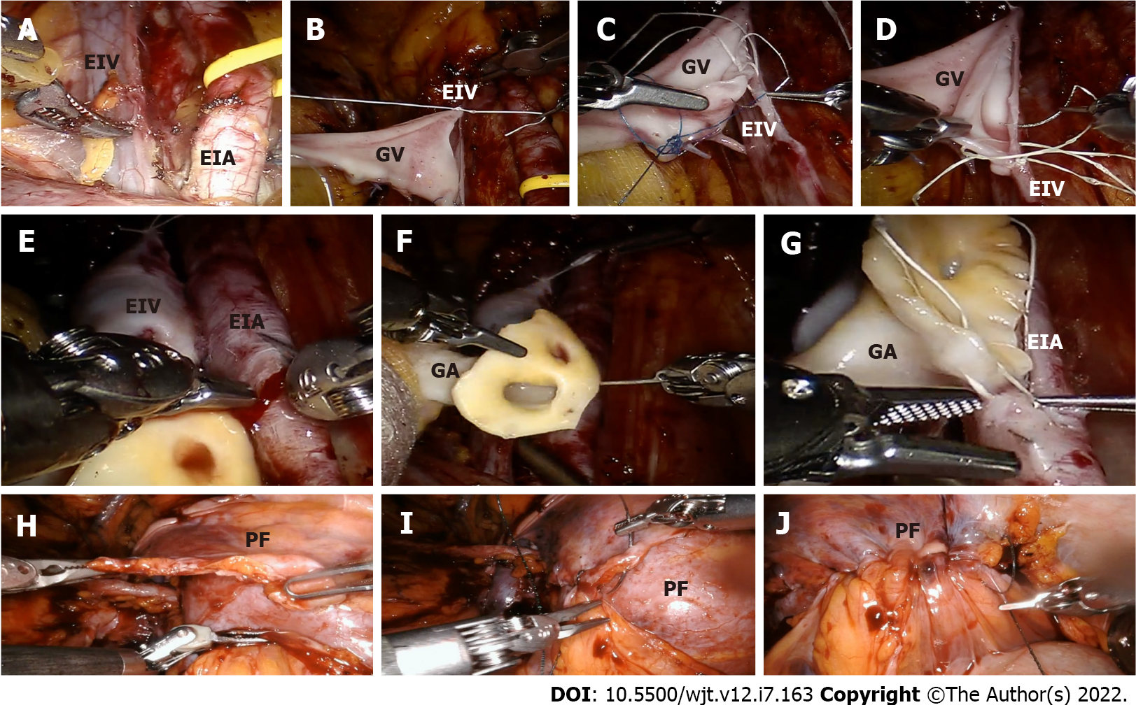
Figure 2 Intraoperative snapshots showing the main phases of isolation of the vascular and uretero-vesical anastomoses during robot-assisted kidney transplantation from deceased donors.
A: After skeletonization of external iliac vessels, the surgeon created an extraperitoneal pouch over the psoas muscle to allocate the graft after completion of the vascular anastomoses. A distal bulldog clamp followed by a proximal clamp was placed on the external iliac vein; B-D: A longitudinal venotomy with cold scissors was performed, and an end-to-side anastomosis between the graft renal vein and the external iliac vein was completed in an end-to-side fashion using a running suture; E: The previously placed bulldog clamps were released and positioned proximally and then distally on the external iliac artery. After the realization of the arteriotomy; F and G: A continuous end-to-side anastomosis was performed between the external iliac and the graft artery. Subsequently, the uretero-vesical anastomosis was performed according to a modified Lich-Gregoire technique; H-J: The graft is allocated in the previously prepared extraperitoneal pouch by reapproximating the two peritoneal flaps prepared at the beginning of the procedure. EIA: External iliac artery; EIV: External iliac vein; GA: Graft artery; GV: Graft vein; PF: Peritoneal flap.
- Citation: Li Marzi V, Pecoraro A, Gallo ML, Caroti L, Peris A, Vignolini G, Serni S, Campi R. Robot-assisted kidney transplantation: Is it getting ready for prime time? World J Transplant 2022; 12(7): 163-174
- URL: https://www.wjgnet.com/2220-3230/full/v12/i7/163.htm
- DOI: https://dx.doi.org/10.5500/wjt.v12.i7.163


