Copyright
©The Author(s) 2022.
World J Transplant. Jan 18, 2022; 12(1): 15-20
Published online Jan 18, 2022. doi: 10.5500/wjt.v12.i1.15
Published online Jan 18, 2022. doi: 10.5500/wjt.v12.i1.15
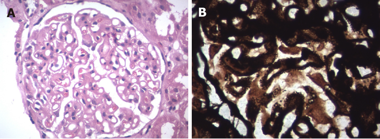
Figure 1 Light microscopy.
A: Haematoxylin and eosin staining (40 × magnification) showed diffuse thickening of the glomerular basement membrane; B: Periodic acid-Schiff silver methenamine stain (100 × magnification) showed membrane thickening.
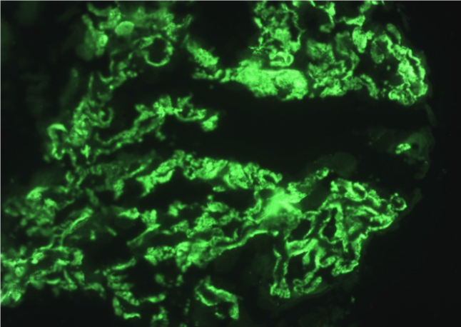
Figure 2 Immunofluorescence microscopy showed granular IgG deposits in the glomerular basement membrane.
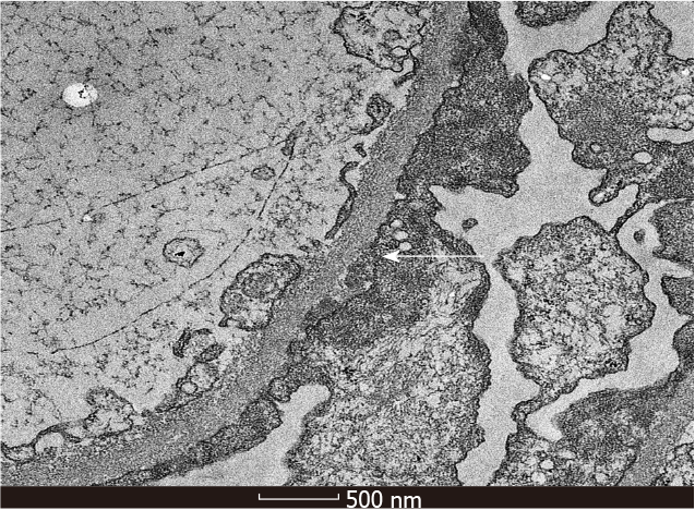
Figure 3 Electron microscopy showed subepithelial electron dense deposits (× 13500).
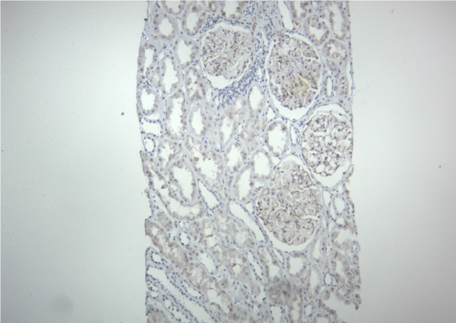
Figure 4 Immunohistochemistry (10 × magnification) was negative for C4d stain.
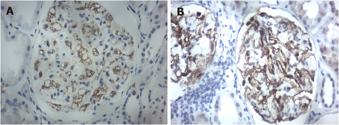
Figure 5 Immunohistochemistry staining (40 × magnification).
A: IgG4 was negative; B: IgG1 was positive.
- Citation: Darji PI, Patel HA, Darji BP, Sharma A, Halawa A. Is de novo membranous nephropathy suggestive of alloimmunity in renal transplantation? A case report. World J Transplant 2022; 12(1): 15-20
- URL: https://www.wjgnet.com/2220-3230/full/v12/i1/15.htm
- DOI: https://dx.doi.org/10.5500/wjt.v12.i1.15


