Copyright
©The Author(s) 2015.
World J Rheumatol. Jul 12, 2015; 5(2): 69-81
Published online Jul 12, 2015. doi: 10.5499/wjr.v5.i2.69
Published online Jul 12, 2015. doi: 10.5499/wjr.v5.i2.69
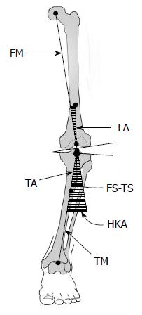
Figure 1 Diagram of a varus knee illustrating the mechanical and anatomic axes and angles.
The FS-TS angle is 4° to 6° valgus compared to the HKA angle. (Modified from Cooke and Sled[46]). FM: Femoral mechanical axis; TM: Tibial mechanical axis; FA: Femoral anatomic axis; TA: Tibial anatomic axis; HKA: Hip-knee-ankle angle (mechanical angle); FS-TS: Femoral shaft-tibial shaft angle (anatomic angle).
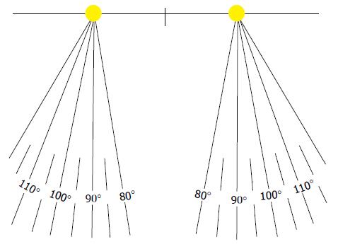
Figure 2 Calibrated template, used to position feet and to reliably measure lower extremity rotation.
(Modified from Orthopedic Alignment and Imaging Systems, Inc.)
Figure 3 Knee radiograph assessed with representative global, composite and individual feature osteoarthritis grading scales.
The knee is in neutral rotation and slight varus alignment. The medial tibiofemoral compartment is most-affected. OA: Osteoarthritis.
- Citation: Sheehy L, Cooke TDV. Radiographic assessment of leg alignment and grading of knee osteoarthritis: A critical review. World J Rheumatol 2015; 5(2): 69-81
- URL: https://www.wjgnet.com/2220-3214/full/v5/i2/69.htm
- DOI: https://dx.doi.org/10.5499/wjr.v5.i2.69


