Copyright
©2011 Baishideng Publishing Group Co.
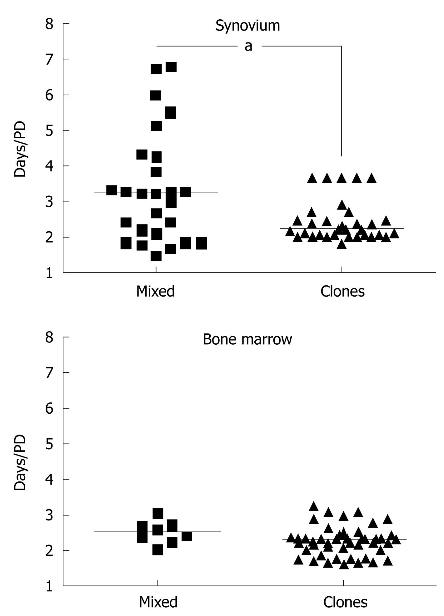
Figure 1 Growth rates of mesenchymal stem cells derived from human synovium and bone marrow.
Faster growth rates of clonal synovial mesenchymal stem cell (MSC) cultures compared to standard polyclonal cultures (mixed with synovial fibroblasts; n = 26 donors, aP < 0.05). Bone marrow MSCs are used as the control (n = 9 donors, P = 0.06). Median values are shown as horizontal bars. PD: Population doubling.
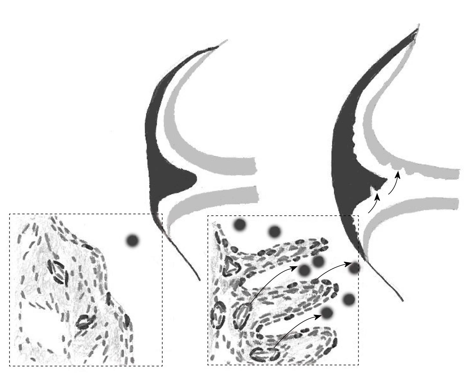
Figure 2 Schematic of the potential involvement of synovial mesenchymal stem cells in cartilage and meniscal repair following injury.
Left: Normal joint, only few mesenchymal stem cells (MSCs) escape into the fluid; Right: Cartilage/meniscal injury leads to MSC to egress into fluid and to traffic along chemotactic gradients emanating from injury-induced signaling centers. Proposed topographical niches of synovial MSCs are shown in the inserts and include synovial lining and perivascular distribution in sublining regions.
-
Citation: Jones E, McGonagle D. Synovial mesenchymal stem cells
in vivo : Potential key players for joint regeneration. World J Rheumatol 2011; 1(1): 4-11 - URL: https://www.wjgnet.com/2220-3214/full/v1/i1/4.htm
- DOI: https://dx.doi.org/10.5499/wjr.v1.i1.4


