Copyright
©The Author(s) 2024.
World J Psychiatry. May 19, 2024; 14(5): 624-634
Published online May 19, 2024. doi: 10.5498/wjp.v14.i5.624
Published online May 19, 2024. doi: 10.5498/wjp.v14.i5.624
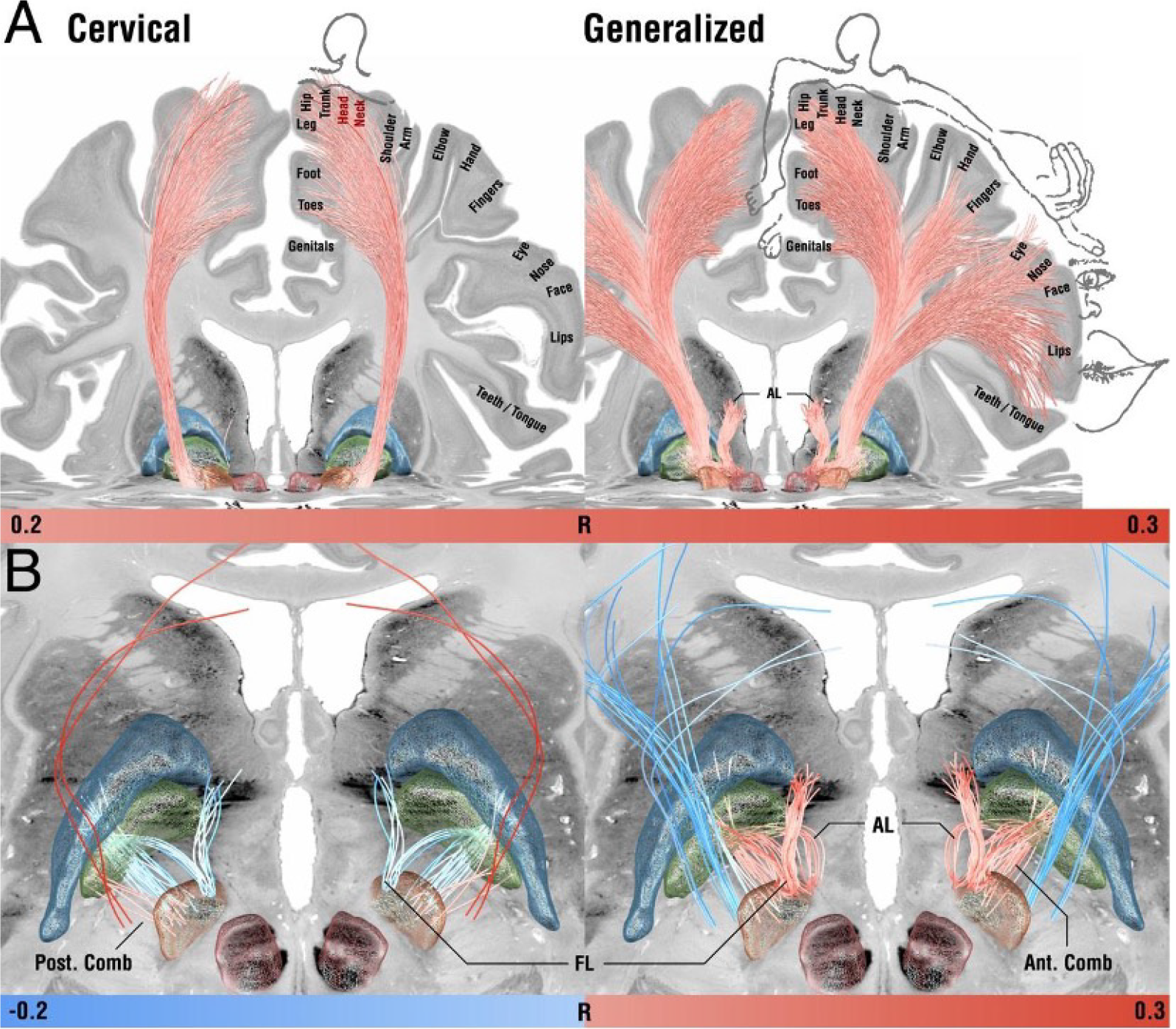
Figure 1 Tracts associated with optimal outcome for patients with cervical (left) and generalized (right) dystonia[85].
A: On a broader scale (slightly lower threshold), modulation of corticofugal tracts from the somatomotor head and neck region was associated with optimal outcomes in cervical dystonia, while tracts from the whole somatotopical domain were associated with generalized dystonia; B: On a localized level (slightly higher threshold), in cervical dystonia, striatopallidofugal tracts of the posterior comb system were associated with optimal outcomes. In contrast, fibers from the fasciculus lenticularis were negatively associated. In generalized dystonia, both pallidothalamic bundles (ansa and fasciculus lenticularis) were associated with optimal outcomes, as was a more anterior portion of the comb system; C: Across the cervical and generalized cohorts, the degree of how fittingly the identified networks were modulated by each patient’s E-field correlated with clinical improvements. While these correlation analyses are of circular nature, a permutation statistic (bottom) showed superior model fits for unpermuted vs permuted improvement values. Citation: Horn A, Reich MM, Ewert S, Li N, Al-Fatly B, Lange F, Roothans J, Oxenford S, Horn I, Paschen S, Runge J, Wodarg F, Witt K, Nickl RC, Wittstock M, Schneider GH, Mahlknecht P, Poewe W, Eisner W, Helmers AK, Matthies C, Krauss JK, Deuschl G, Volkmann J, Kühn AA. Optimal deep brain stimulation sites and networks for cervical vs. generalized dystonia. Proc Natl Acad Sci U S A 2022; 119: e2114985119. Copyright© The Authors 2022. Published by National Academy of Sciences of the United States (Supplementary material).
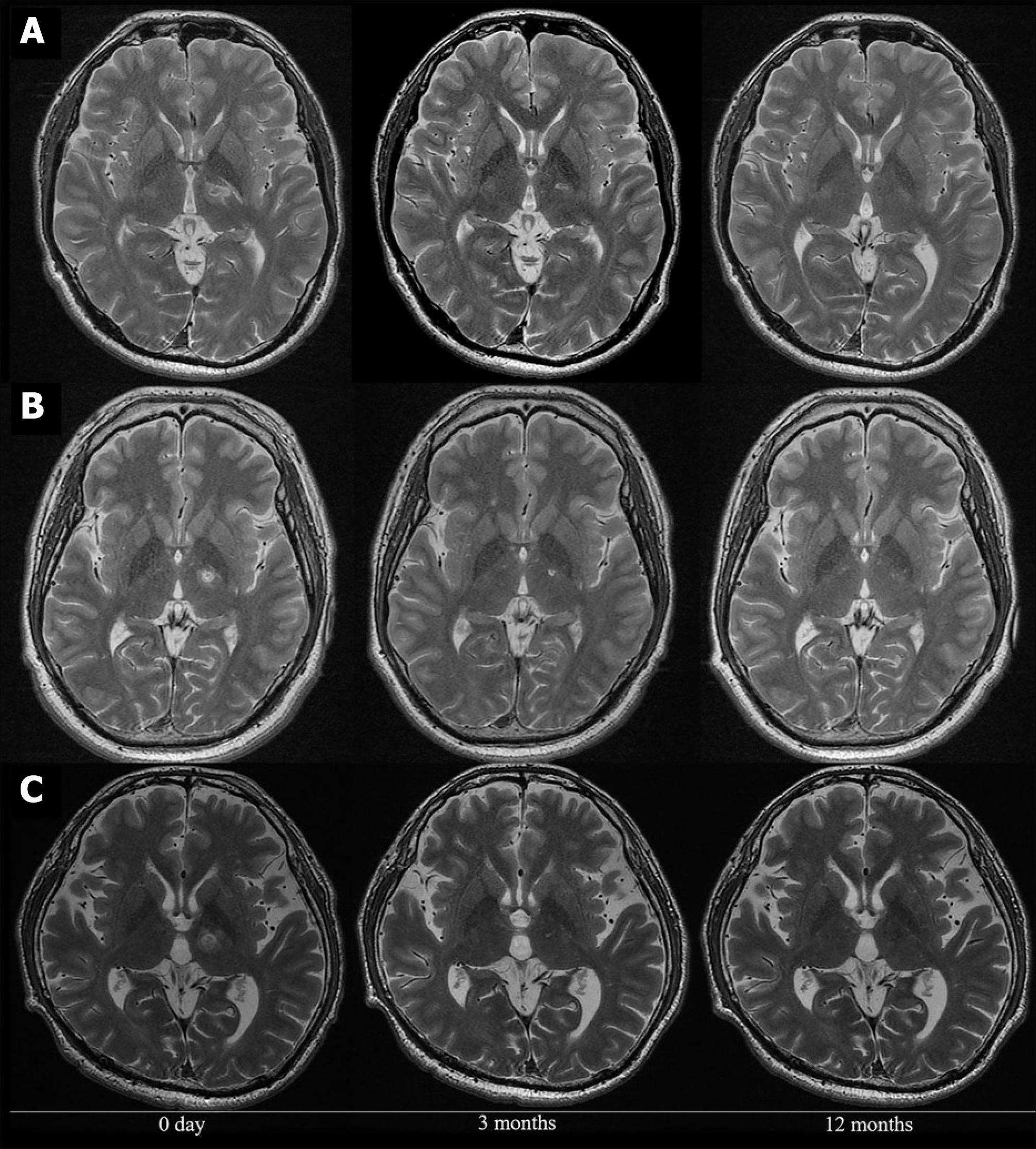
Figure 2 Representative magnetic resonance images obtained before and after treatment[86].
A and B: The lesion unexpectedly encroached on the posterior limb of the internal capsule (case 4: A; case 7: B); C: The precise lesion on the intended target. Citation: Horisawa S, Yamaguchi T, Abe K, Hori H, Fukui A, Iijima M, Sumi M, Hodotsuka K, Konishi Y, Kawamata T, Taira T. Magnetic Resonance-Guided Focused Ultrasound Thalamotomy for Focal Hand Dystonia: A Pilot Study. Mov Disord 2021; 36: 1955-1959. Copyright© The Authors 2021. Published by Movement Disorders published by John Wiley & Sons, Inc. (Supplementary material).
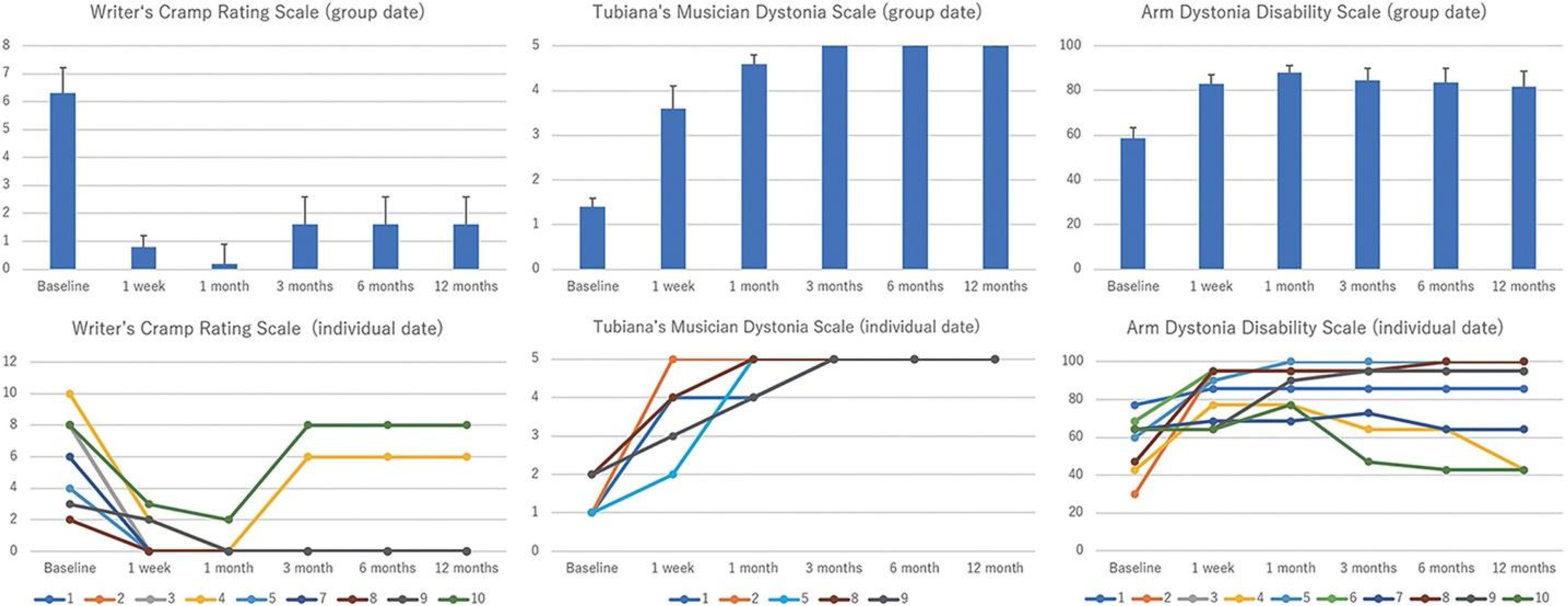
Figure 3 Changes in primary and secondary clinical endpoints during the study period[86].
Primary clinical endpoints were the scores of the Writer’s Cramp Rating Scale (WCRS; ranging from 0 to 30, with higher scores indicating greater severity) and Tubiana Musician’s Dystonia Scale (TMDS; ranging from 1 to 5, with lower scores indicating greater severity), which evaluate dystonia severity. Secondary clinical endpoint was the score of the Arm Dystonia Disability Scale (ADDS; ranging from 0% to 100%, with lower scores indicating greater disability) to evaluate dystonia disability. Significant improvements were observed in the WCRS, TMDS, and ADDS scores throughout the study period in group data (upper row). Individual data are shown in the lower row. Citation: Horisawa S, Yamaguchi T, Abe K, Hori H, Fukui A, Iijima M, Sumi M, Hodotsuka K, Konishi Y, Kawamata T, Taira T. Magnetic Resonance-Guided Focused Ultrasound Thalamotomy for Focal Hand Dystonia: A Pilot Study. Mov Disord 2021; 36: 1955-1959. Copyright© The Authors 2021. Published by Movement Disorders published by John Wiley & Sons, Inc. (Supplementary material).
- Citation: Mohamed AA, Faragalla S, Khan A, Flynn G, Rainone G, Johansen PM, Lucke-Wold B. Neurosurgical and pharmacological management of dystonia. World J Psychiatry 2024; 14(5): 624-634
- URL: https://www.wjgnet.com/2220-3206/full/v14/i5/624.htm
- DOI: https://dx.doi.org/10.5498/wjp.v14.i5.624


