Copyright
©The Author(s) 2024.
World J Psychiatry. Mar 19, 2024; 14(3): 456-466
Published online Mar 19, 2024. doi: 10.5498/wjp.v14.i3.456
Published online Mar 19, 2024. doi: 10.5498/wjp.v14.i3.456
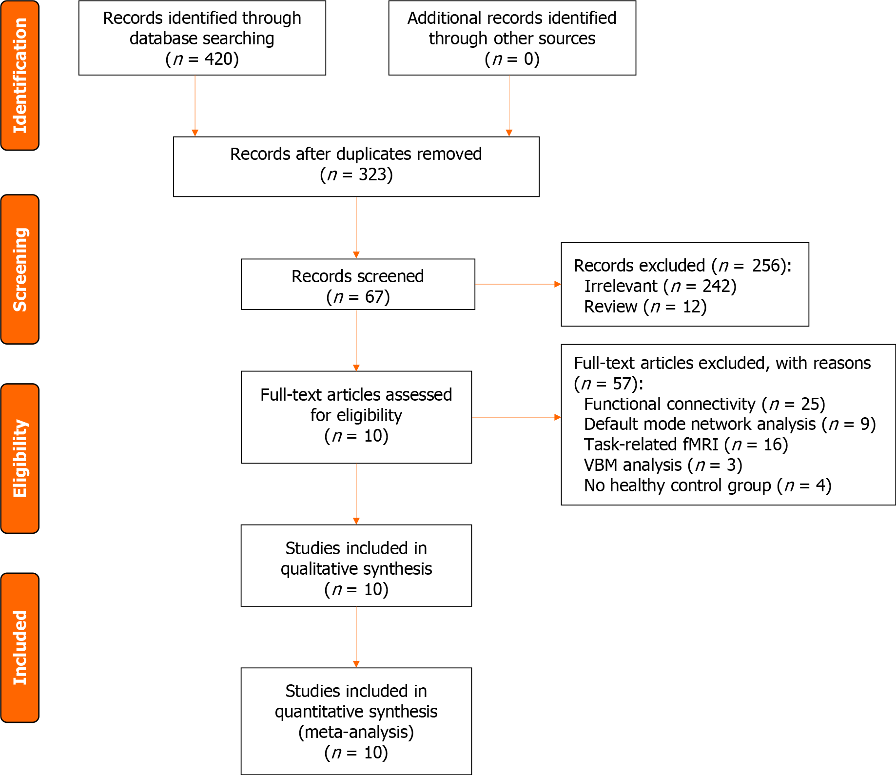
Figure 1 Flow chart of the study selection strategy.
VBM: Voxel-based morphometry; fMRI: Functional magnetic resonance imaging.
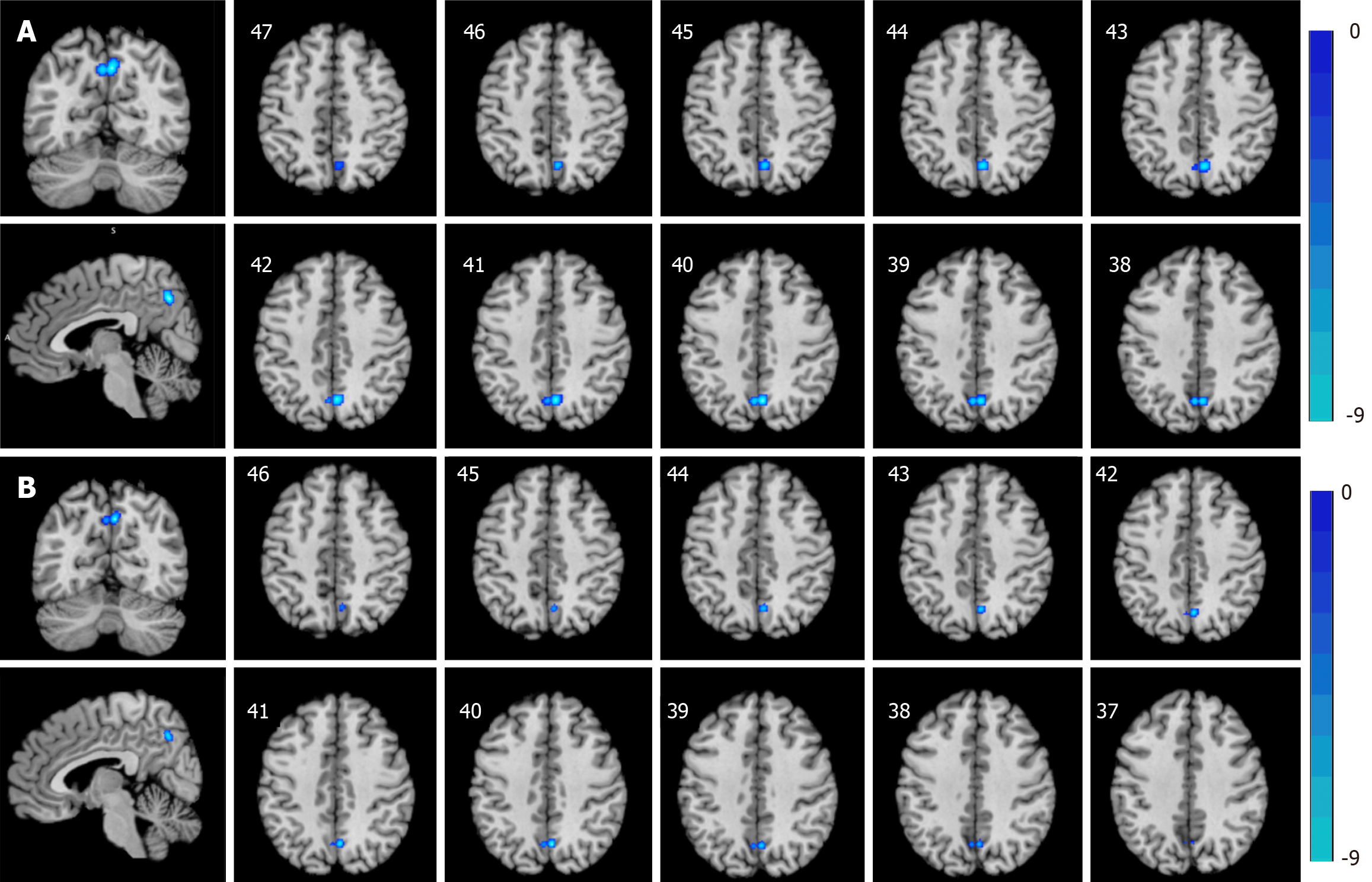
Figure 2 Schematic construction of brain areas with decreased activity in adolescents with major depressive disorder relative to healthy controls (cluster-level FWE correction at P < 0.
05). A: Regional homogeneity and amplitude of low-frequency fluctuations (ALFF)/fractional ALFF methods; B: The ALFF method.
- Citation: Ding H, Zhang Q, Shu YP, Tian B, Peng J, Hou YZ, Wu G, Lin LY, Li JL. Vulnerable brain regions in adolescent major depressive disorder: A resting-state functional magnetic resonance imaging activation likelihood estimation meta-analysis. World J Psychiatry 2024; 14(3): 456-466
- URL: https://www.wjgnet.com/2220-3206/full/v14/i3/456.htm
- DOI: https://dx.doi.org/10.5498/wjp.v14.i3.456


