Copyright
©The Author(s) 2023.
World J Psychiatry. Sep 19, 2023; 13(9): 620-629
Published online Sep 19, 2023. doi: 10.5498/wjp.v13.i9.620
Published online Sep 19, 2023. doi: 10.5498/wjp.v13.i9.620
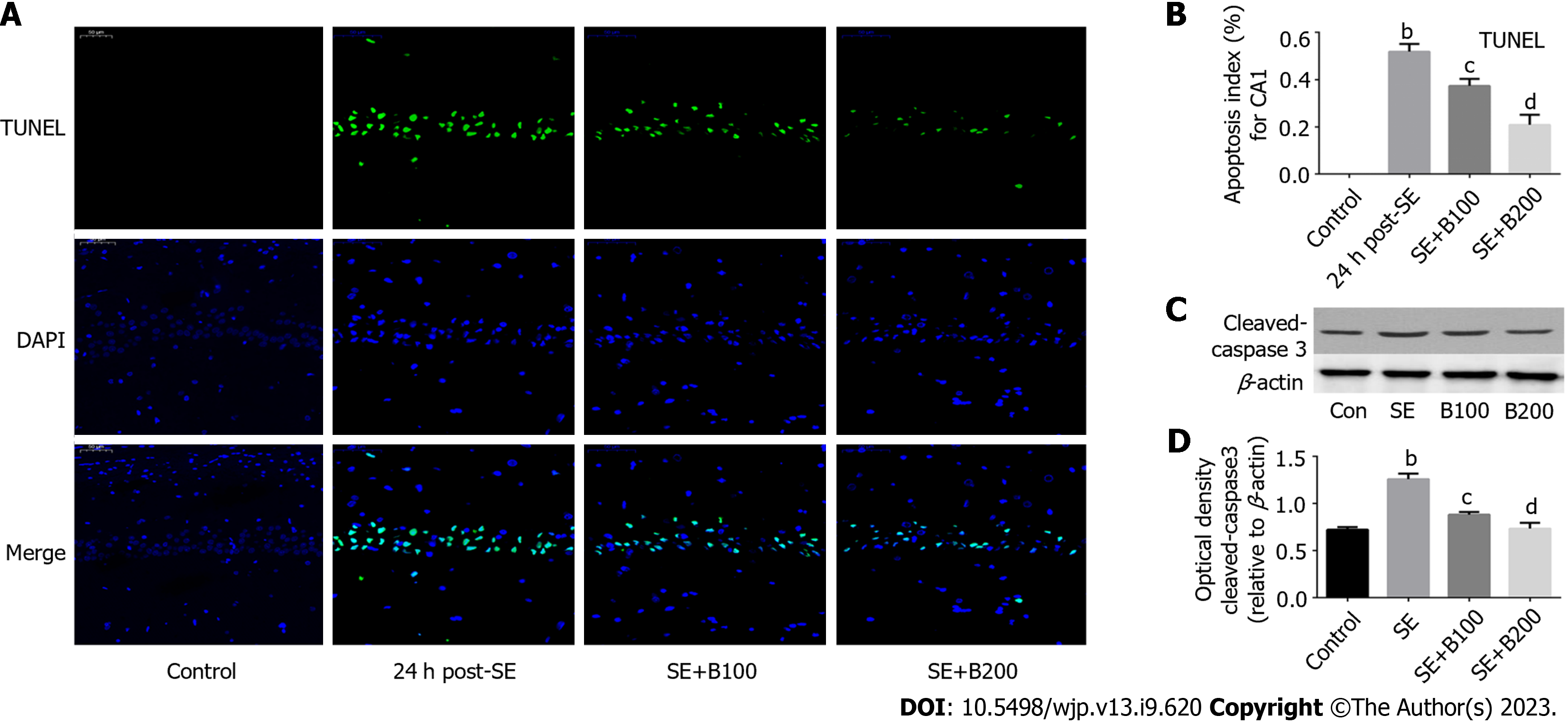
Figure 1 Baicalin ameliorated status epilepticus-induced neuronal apoptosis.
A: Transferase dUTP nick end labeling staining was performed; B: Neuronal apoptosis was analyzed; C: Western blotting was performed; D: Semi-quantitative analysis of cleaved caspase-3 (n = 6). bP < 0.01 compared to the control group; cP < 0.05 and dP < 0.01 compared to the status epilepticus group. Scale bar = 50 μm. SE: Status epilepticus; TUNEL: Transferase dUTP nick end labeling.
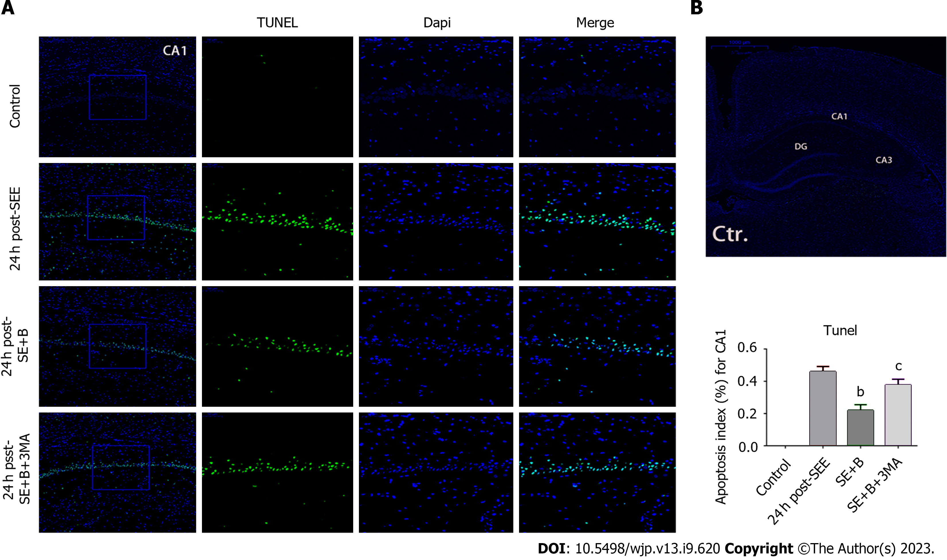
Figure 2 Baicalin ameliorated status epilepticus-induced neuronal apoptosis.
A: Transferase dUTP nick end labeling staining was performed; B: Neuronal apoptosis was analyzed. Baicalin protects the hippocampus from apoptosis following status epilepticus (SE), and 3-Methyladenine reverses Baicalin-induced neuroprotection in hippocampal neurons. Data are represented as mean ± SD. (n = 5), bP < 0.01 vs the SE group; cP < 0.05 vs the SE + B group. Scale bar = 50 μm. SE: Status epilepticus; TUNEL: Transferase dUTP nick end labeling; 3-MA: 3-Methyladenine.
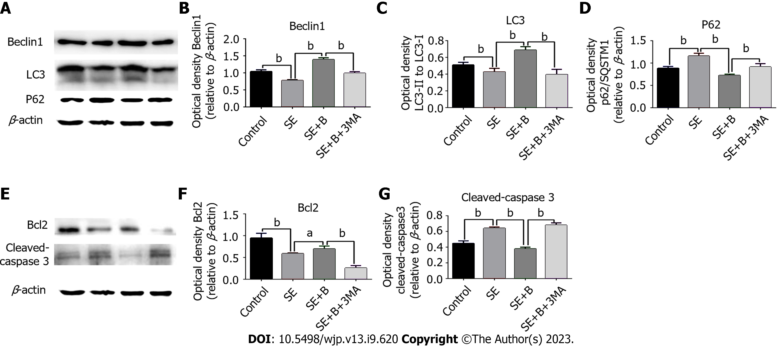
Figure 3 The autophagy markers (p62/SQSTM1, Beclin 1, and LC3) and apoptotic pathway markers (cleaved caspase-3 and Bcl-2) were measured using western blotting.
A: The autophagy markers (p62/SQSTM1, Beclin 1, and LC3) were measured using western blotting; B: The expressions of Beclin 1; C: The expressions of LC3; D: The expressions of p62; E: The apoptotic pathway markers (cleaved caspase-3 and Bcl-2) were measured using western blotting; F: The expressions of Bcl-2; G: Cleaved caspase-3 were analyzed. n = 6. aP < 0.05 and bP < 0.01. SE: Status epilepticus; 3-MA: 3-Methyladenine.
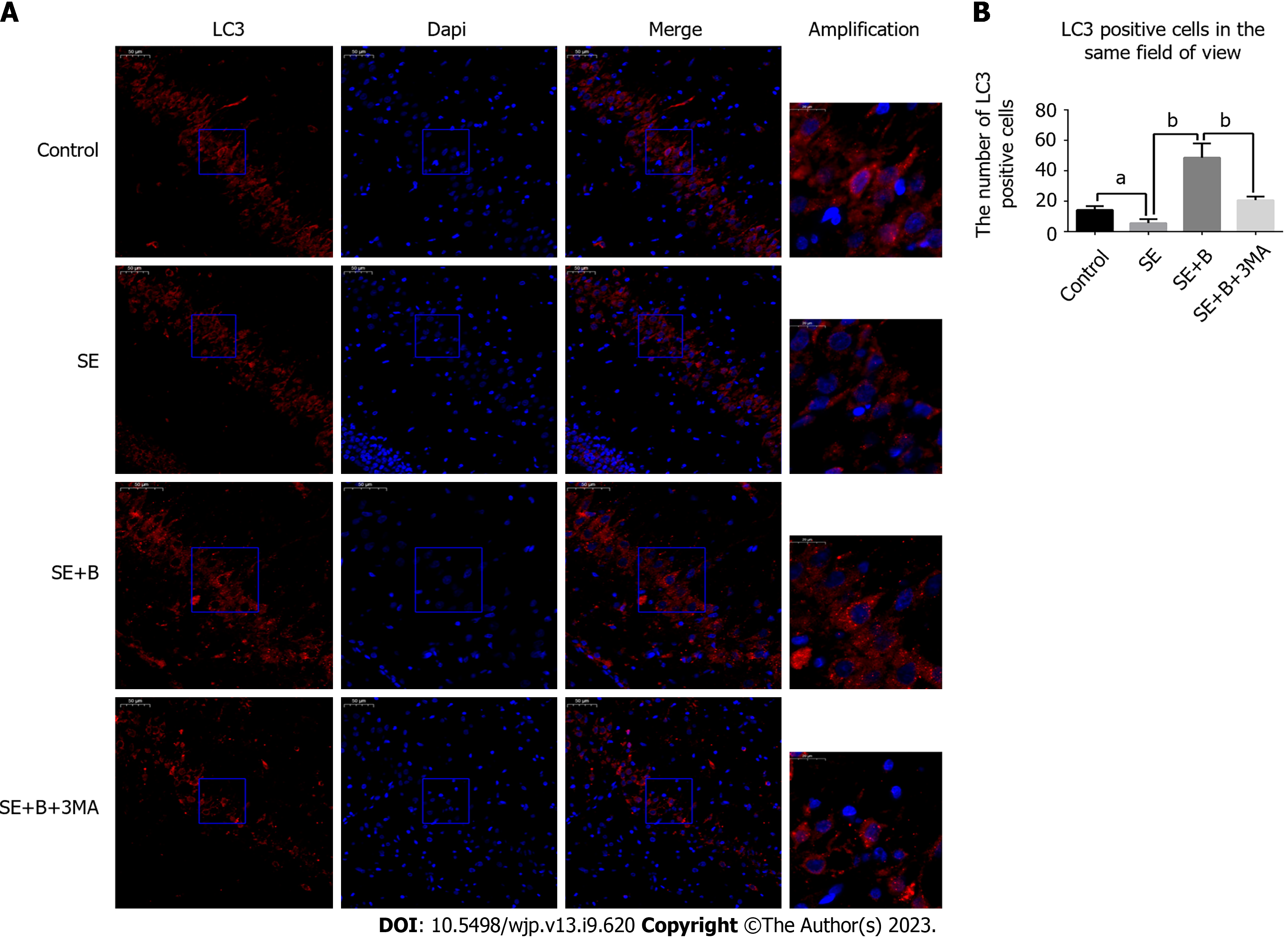
Figure 4 The number of LC3-II-positive neurons was partially decreased by SE and increased by Baicalin.
3-Methyladenine reversed the Baicalin-induced alteration. A: Representative immunofluorescence staining; B: Positive neuronal cells were analyzed. n = 6. aP < 0.05 and bP < 0.01 vs the relevant group. The number of LC3-II-positive cells/0.5-mm-long subfield of the hippocampus under a light microscope was regarded as the numerical value (scale bar = 50 μm). SE: Status epilepticus; 3-MA: 3-Methyladenine.
- Citation: Yang B, Wen HY, Liang RS, Lu TM, Zhu ZY, Wang CH. Hippocampus protection from apoptosis by Baicalin in a LiCl-pilocarpine-induced rat status epilepticus model through autophagy activation. World J Psychiatry 2023; 13(9): 620-629
- URL: https://www.wjgnet.com/2220-3206/full/v13/i9/620.htm
- DOI: https://dx.doi.org/10.5498/wjp.v13.i9.620


