Copyright
©The Author(s) 2016.
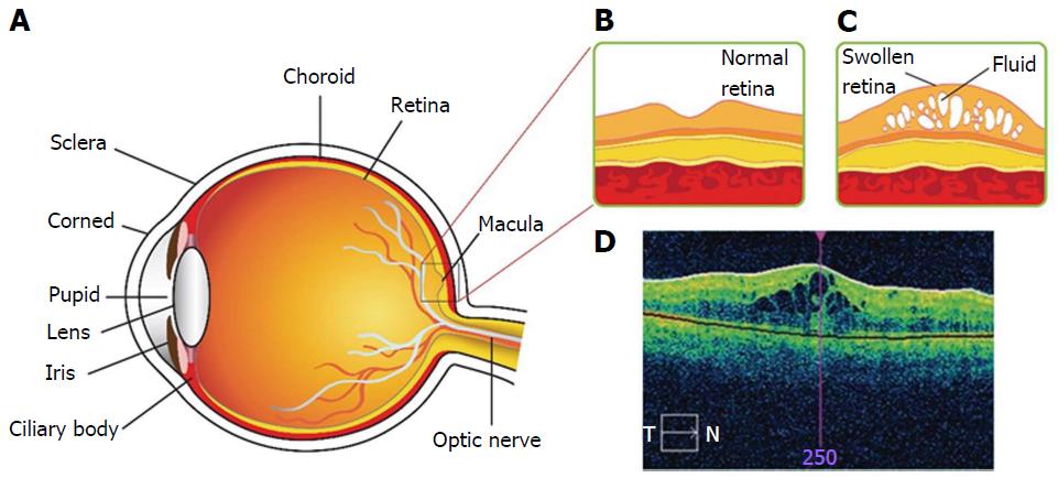
Figure 1 Diabetic macular edema at disease state.
A: Structure of human eye; B: Expanded representation of macula region for normal eye; C: Expanded representation of macula region for diabetic macular edema (DME); D: Optical coherence tomography image for DME.
- Citation: Trinh HM, Joseph M, Cholkar K, Pal D, Mitra AK. Novel strategies for the treatment of diabetic macular edema. World J Pharmacol 2016; 5(1): 1-14
- URL: https://www.wjgnet.com/2220-3192/full/v5/i1/1.htm
- DOI: https://dx.doi.org/10.5497/wjp.v5.i1.1


