Copyright
©The Author(s) 2015.
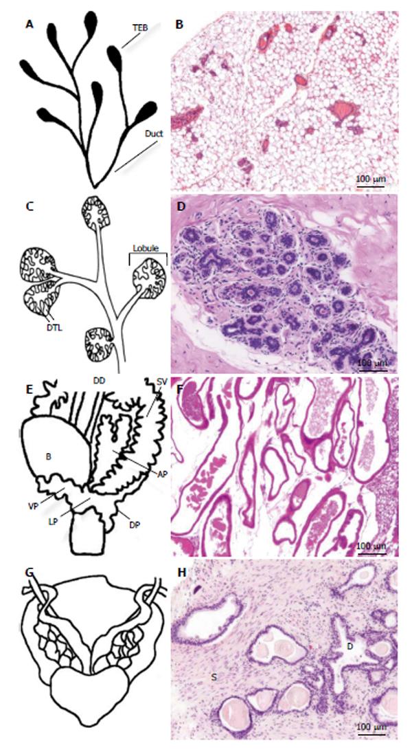
Figure 1 Anatomical and histological comparison of mouse and human mammary gland (A-D) or prostate (E-H).
A: Schematic representation of pubertal mouse mammary tree ducts, which end in club shaped terminal end buds (TEBs); B: Hematoxylin & eosin (H&E) stained section of mouse breast tissue, showing ducts imbedded in a stroma composed of adipose tissue; C: Human mature nulliparous terminal ductal lobular unit, 30-50 ductules (DTL) are present in each lobule; D: H&E stained section of human mammary gland showing a terminal ductal lobular unit comprised of ducts and acini in a fibrous connective tissue stroma; E: Mouse prostate surrounds urethra and has distinct lobes: ventral lobe (VP), dorsal lobe (DP) and lateral lobe (LP); F: H&E stained section of mouse ventral prostate; G: Human prostate is a nut shaped glad which also surrounds the urethra; H: The proportion of stroma in human prostate is larger compared with mouse prostate, H&E staining shows secreting ducts (D) and stroma (S). B: Bladder; DD: Ductus deferens; SV: Seminal vesicle; AP: Anterior prostate.
- Citation: Valta M, Fagerlund K, Suominen M, Halleen J, Tuomela J. Importance of microenvironment in preclinical models of breast and prostate cancer. World J Pharmacol 2015; 4(1): 47-57
- URL: https://www.wjgnet.com/2220-3192/full/v4/i1/47.htm
- DOI: https://dx.doi.org/10.5497/wjp.v4.i1.47


