Copyright
©The Author(s) 2015.
World J Clin Infect Dis. Feb 25, 2015; 5(1): 11-13
Published online Feb 25, 2015. doi: 10.5495/wjcid.v5.i1.11
Published online Feb 25, 2015. doi: 10.5495/wjcid.v5.i1.11
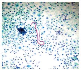
Figure 1 Ascitic fluid-Smear shows Microfilaria.
Background shows inflammatory cells (Pap stain 100 ×).
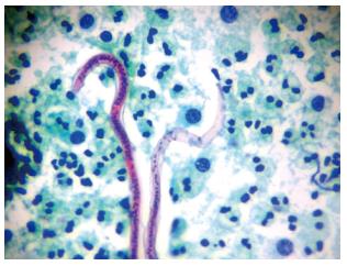
Figure 2 Microfilaria with nuclei.
Note that the tail portion is devoid of the nuclei (pap 400 ×).
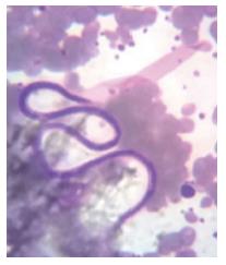
Figure 3 Microfilaria in peripheral smear.
Sample collected at 2 am.
- Citation: Shah KS, Bhate PA, Solanke D, Pandey V, Ingle MA, Kane SV, Sawant P. Non chylous filarial ascites: A rare case report. World J Clin Infect Dis 2015; 5(1): 11-13
- URL: https://www.wjgnet.com/2220-3176/full/v5/i1/11.htm
- DOI: https://dx.doi.org/10.5495/wjcid.v5.i1.11


