Copyright
©2014 Baishideng Publishing Group Co.
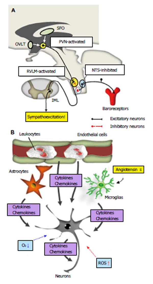
Figure 1 Abnormal inflammatory responses in the cardiovascular center and neurogenic hypertension.
A: Sympathetic pre-motor neurons controlling vasomotor sympathetic nerve activity are located primarily in the rostral ventrolateral medulla (RVLM). These neurons receive numerous excitatory and inhibitory inputs from other areas of the brain, such as the hypothalamus and parts of the medulla oblongata. Over-activation of hypothalamic paraventricular nucleus (PVN) and RVLM neurons and/or attenuated nucleus tractus solitarius (NTS) responses to baroreceptor inputs are probable mechanisms of neurogenic hypertension (see text). Abnormal inflammatory responses may contribute to altered neuronal functions in the cardiovascular center; B: Inflammatory molecules released from vascular endothelial cells, leukocytes, astrocytes, microglial cells and neurons may affect neuronal functions in the cardiovascular center and induce sympathoexcitation. Hypoxia and ROS production induced by abnormal inflammatory responses may also affect neuronal functions (See text). OVLT: Organum vasculosum of the lamina terminalis; SFO: Subfornical organ; IML: Intermediolateral cell column; ROS: Reactive oxygen species.
- Citation: Waki H, Gouraud SS. Brain inflammation in neurogenic hypertension. World J Hypertens 2014; 4(1): 1-6
- URL: https://www.wjgnet.com/2220-3168/full/v4/i1/1.htm
- DOI: https://dx.doi.org/10.5494/wjh.v4.i1.1


