Basic Study Open Access
Copyright ©The Author(s) 2024. Published by Baishideng Publishing Group Inc. All rights reserved.
World J Exp Med. Jun 20, 2024; 14(2): 90374
Published online Jun 20, 2024. doi: 10.5493/wjem.v14.i2.90374
Expression levels of KATP channel subunits and morphological changes in the mouse liver after exposure to radiation
Ming Zhou, Hideo Akashi, Ryoji Suzuki, Yoshio Bando, Department of Anatomy, Akita University Graduate School of Medicine, Akita 010-8543, Japan
Tao-Sheng Li, Department of Stem Cell Biology, Atomic Bomb Disease Institute, Nagasaki University, Nagasaki 852-8523, Japan
Hiroshi Abe, Sendai Old Age Refresh Station, A Long-term Care Health Facility, Sendai 981-1105, Japan
ORCID number: Ming Zhou (0000-0001-8775-9738); Tao-Sheng Li (0000-0002-7653-8873); Hiroshi Abe (0000-0002-4683-4487); Hideo Akashi (0000-0001-8520-0252); Ryoji Suzuki (0000-0003-4526-7406); Yoshio Bando (0000-0002-2466-5248).
Co-corresponding authors: Ming Zhou and Tao-Sheng Li.
Author contributions: Zhou M performed the main experiments, collected the data, and wrote the manuscript; Akashi H and Suzuki R participated in the treatment of animals; Li TS designed the program and experiment; Li TS, Abe H, and Bando Y supervised the investigation and approved the manuscript for publication. This study was a collaboration between Zhou M and Li TS. Both authors applied for and obtained the funds for this research project, and have made indispensable contributions to completion of this research and manuscript preparation as the co-corresponding authors. This collaboration between Zhou M and Li TS is crucial for the publication of this manuscript.
Supported by the Program of the Network-type Joint Usage/Research Center for Radiation Disaster Medical Science of Hiroshima University, Nagasaki University.
Institutional review board statement: This study was reviewed and approved by the review board of Atomic Bomb Disease Institute, Nagasaki University.
Institutional animal care and use committee statement: This study was approved by the Nagasaki University Japan’s Institutional Animal Care and Use Committee and conducted following the Committee’s protocols. All animal procedures followed the institutional and national guidelines.
Informed consent statement: This study has not involved human subjects.
Conflict-of-interest statement: All authors have no conflicts of interest to disclose.
Data sharing statement: No sharing data with this research.
ARRIVE guidelines statement: The authors have read the ARRIVE guidelines, and the manuscript was prepared and revised according to the ARRIVE guidelines.
Open-Access: This article is an open-access article that was selected by an in-house editor and fully peer-reviewed by external reviewers. It is distributed in accordance with the Creative Commons Attribution NonCommercial (CC BY-NC 4.0) license, which permits others to distribute, remix, adapt, build upon this work non-commercially, and license their derivative works on different terms, provided the original work is properly cited and the use is non-commercial. See: https://creativecommons.org/Licenses/by-nc/4.0/
Corresponding author: Ming Zhou, MD, PhD, Assistant Professor, Department of Anatomy, Akita University Graduate School of Medicine, 1-1-1 Hondo, Akita 010-8543, Japan. m.ming.zhou99@gmail.com
Received: December 7, 2023
Revised: January 28, 2024
Accepted: March 27, 2024
Published online: June 20, 2024

Abstract
BACKGROUND

ATP sensitive K+ (KATP) channels are ubiquitously distributed in various of cells and tissues, including the liver. They play a role in the pathogenesis of myocardial and liver ischemia.

AIM

To evaluate the radiation-induced changes in the expression of KATP channel subunits in the mouse liver to understand the potential role of KATP channels in radiation injury.

METHODS

Adult C57BL/6 mice were randomly exposed to γ-rays at 0 Gy (control, n = 2), 0.2 Gy (n = 6), 1 Gy (n = 6), or 5 Gy (n = 6). The livers were removed 3 and 24 h after radiation exposure. Hematoxylin and eosin staining was used for morphological observation; immunohistochemical staining was applied to determine the expression of KATP channel subunits in the liver tissue.

RESULTS

Compared with the control group, the livers exposed to 0.2 Gy γ-ray showed an initial increase in the expression of Kir6.1 at 3 h, followed by recovery at 24 h after exposure. Exposure to a high dose of 5.0 Gy resulted in decreased expression of Kir6.1 and increased expression of SUR2B at 24 h. However, the expression of Kir6.2, SUR1, or SUR2A had no remarkable changes at 3 and 24 h after exposure to any of these doses.

CONCLUSION

The expression levels of Kir6.1 and SUR2B in mouse liver changed differently in response to different radiation doses, suggesting a potential role for them in radiation-induced liver injury.

Key Words: Radiation exposure, ATP-sensitive K+ channel, Mouse, Liver

Core Tip: ATP-sensitive K+ (KATP) channels are ubiquitously distributed in various cell types and tissues, including the liver; however, their role in the development of radiation-induced liver damage remains unknown. In the current study, the expression of KATP channel subunits in the liver tissue changed dose-dependently in response to radiation exposure, suggesting their potential role in radiation-induced liver injury.


INTRODUCTION

Radiation-induced liver injury is becoming increasingly familiar with the advancements in technology and increased exposure to low levels of radiation during medical procedures and treatment[1]. Ionizing radiation can induce functional and structural changes in the liver, leading to radiation-induced liver diseases[2]. Using radiation therapy for the treatment of hepatic tumors is rapidly increasing; the application of radiation treatment for tumors in the liver, right lower lung, distal esophagus, and whole abdomen is on the increase, resulting in cell damage to the non-tumor compartments of the liver[3,4]. Clinically, radiation-induced liver diseases have drawn particular attention from physicians recently as the main challenge in delivering curative tumors doses[5]. Novel approaches to prevent radiation-induced liver damage during radiation therapy are urgently needed.

ATP-sensitive K+ (KATP) channels are observed in various cells and tissues[6], including pancreatic β cells[7], skeletal and cardiac muscles[8-11], and the brain[12-14], kidneys[15,16], and liver[17,18]. These channels may close or open depending on the concentration of intracellular ATP, playing a protective role during ischemia[19] by coupling changed energy metabolism via membrane potential[20]. KATP channels are formed by two kinds of subunits, pore-forming subunits (Kir6.1 or Kir6.2) and regulatory subunits, such as sulfonylurea receptors (SUR1, SUR2A, and SUR2B), with a hetero-octamer structure of four pore-forming subunits and four regulatory subunits[21,22]. Different subunit compositions show different physiological and pharmacological functions[23,24]. KATP channels in the hypothalamus are involved in glucose homeostasis by controlling glucagon and catecholamines[25]. The liver has diverse functions, such as producing bile, metabolism of ingested nutrients, eliminating waste products, and synthesizing protein; it is directly involved in glucose metabolism[4], which influences the change in intracellular ATP concentrations to which KATP channels are sensitive. KATP channels regulate the proliferation of primary rat hepatocytes[26]. A previous study revealed that KATP channel subunits are widely localized in hepatocytes, hepatic stellate cells, and Kupffer cells[18].

Radiation exposure is generally classified as low (< 0.5 Gy), moderate (0.5-5 Gy), or high (> 5 Gy)[27]. Radiation causes ultrastructural and biochemical changes in hepatocytes, which are known to depend on lipid metabolism[28]. Radiation exposure is one of the causes of hepatic fibrosis after liver injury recovery[29-31]. However, there is no particular information on the role of KATP channel subunits in radiation-induced liver damage. This study investigated the expression of KATP channel subunits in the liver after different irradiation doses.

MATERIALS AND METHODS
Animals

Twenty adult (8-12 wk old) male C57BL/6 mice (CLEA Japan, Inc.) were applied for this study. The study was approved by the Nagasaki University Japan’s Institutional Animal Care and Use Committee and conducted following the Committee’s protocols. All animal procedures followed the institutional and national guidelines. The mice were kept under constant environmental conditions in group cages with a condition of 12-h light/dark cycle and given food and water ad libitum. The mice were randomly separated into four groups: (1) Control, without γ-ray irradiation (n = 2); (2) lower dose (0.2 Gy) exposure (n = 6); (3) medium dose (1 Gy) exposure (n = 6); and (4) high dose (5 Gy) exposure (n = 6).

Radiation exposure

The mice were placed in a special cage for radiation exposure to assess radiation-induced liver injury corresponding to dose dependency. PS-3100SB (Pony Industry Co., Ltd., Osaka, Japan), a γ-ray irradiation system with a Cs source was used. The mice were exposed to a dose of 0, 0.2, 1, or 5 Gy/min. The mice were not anesthetized before irradiation.

Preparation of samples

After radiation exposure, the mice were sacrificed for experiments at 3 and 24 h later. Mice were euthanized via general anesthesia by intraperitoneal injection of pentobarbital (160 mg/kg), followed by perfusion with cold Zamboni’s fixatives through the heart. The liver was quickly collected and further fixed for 6 h in the same fixative, and then put into 30% sucrose over 12 h until it sank down to the bottom. Cryosections were cut (8 to10 µm thick) with a Leica CM1950 cryostat, thaw-mounted on MAS-coated glass slides (Matsunami Industries, Kishiwada, Japan), and kept at -20 ℃ until use.

Histological and immunohistochemical staining

After air-drying for 30 min, several sections were stained with Mayer hematoxylin and eosin (H&E) for morphological observations. The others sections were pre-incubated with 5% normal donkey serum for 30 min, and then incubated with KATP channel subunit antibodies, which were goat anti-human Kir6.1 and Kir6.2 (Santa Cruz Biotechnology), and rabbit anti-rat SUR1, SUR2A, and SUR2B[18] at 1:500 dilution. After washing in PBS three times, 5 min each, the sections were incubated with biotinylated rabbit anti-goat IgG (BA5000; Vector Laboratories, Inc., Burlingame, CA, United States) or biotinylated goat anti-rabbit IgG (BA1000; Vector Laboratories, Inc.) at 1:200 each for 30 min, and subsequently with the ABC complex (Vectastain ABC Kit; Vector Laboratories, Inc.) according to the manufacturer’s instructions. Immunoreactions were carried out with 3,3’-diaminobenzedine in the presence of 0.003% H2O2, and counterstained with Mayer’s hematoxylin.

To show how the pore-forming subunit Kir6.1 co-localizes with the regulatory subunit SUR2B in the liver after radiation, sections were treated with primary goat anti-human Kir6.1 and rabbit anti-rat SUR2B antibodies, each diluted 1:500, for one night (12 h) at room temperature. After being washed three times in PBS for 15 min, the sections were incubated with a mixture of secondary antibodies labeled with Alexa 488 and Alexa 594 (A21432 and A21207; Molecular Probes, Inc., Eugene, OR), each diluted 1:200. Fluorescence images were acquired using an Olympus microscope (BX51; Tokyo, Japan).

RESULTS
Dose dependency of radiation-induced injury in mouse liver

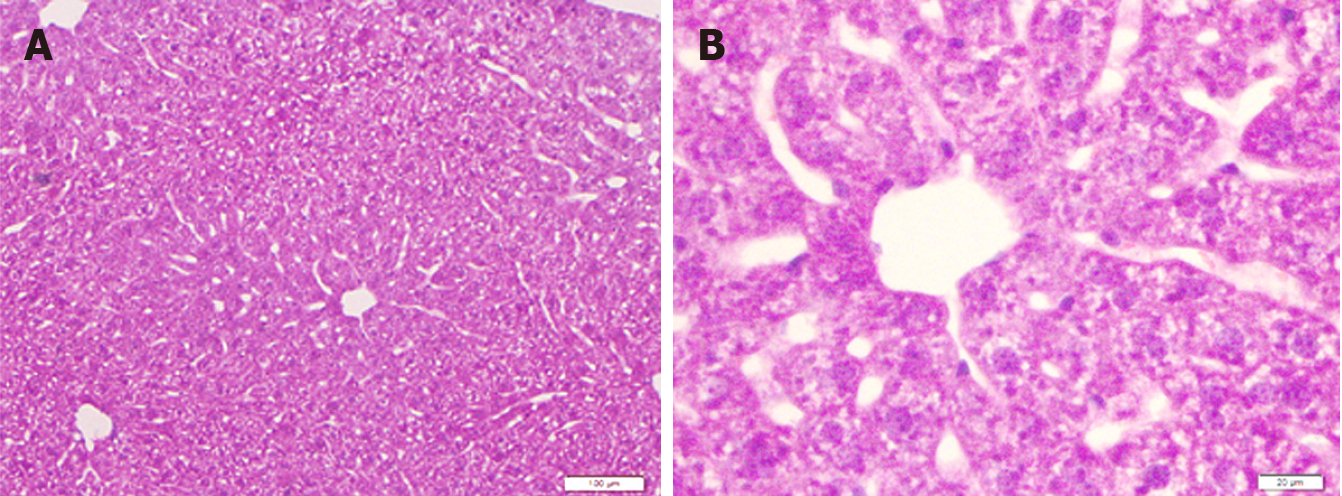
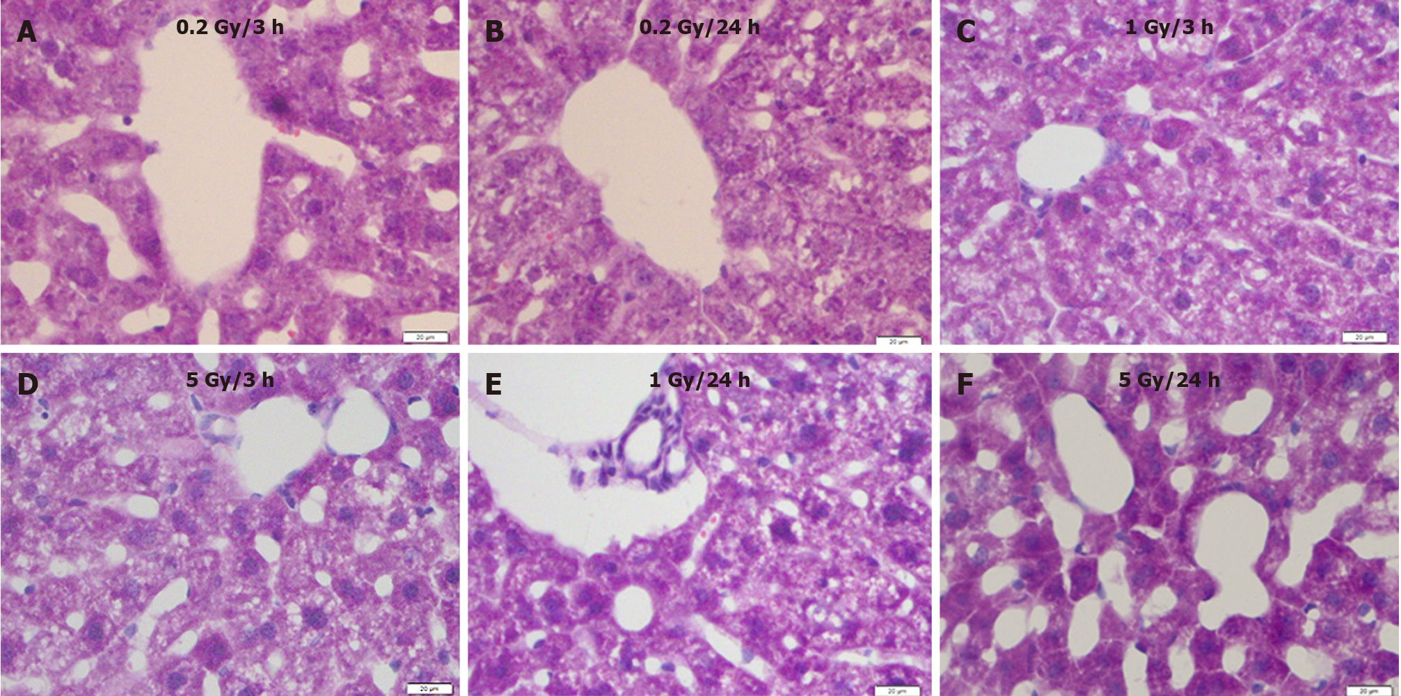
Compared with the control (Figure 1), 0.2 Gy induced hepatocyte atrophy and sinusoid enlargement 3 h after exposure (Figure 2A), which recovered 24 h after irradiation (Figure 2B). Degeneration of hepatocytes, constriction of the hepatocyte cord, and enlarged sinusoids were observed in the 1.0 Gy and 5.0 Gy dose groups 3 h after exposure (Figure 2C and D), with the effects worsening 24 h after γ-ray irradiation (Figure 2E and F).

Figure 1
Figure 1 Liver sections of mice in the control group. A: Low magnification; B: High magnification. H&E staining. Bars: 100 μm (A); 20 μm (B).
Figure 2
Figure 2 Mouse liver sections showing morphological changes with different radiation doses at different time points. A: 3 h after exposure to low dose (0.2 Gy); B: 24 h after exposure to low dose (0.2 Gy); C: 3 h after exposure to medium dose (1 Gy); D: 3 h after exposure to high dose (5 Gy); E: 24 h after exposure to medium dose (1 Gy); F: 24 h after exposure to high dose (5 Gy). H&E staining. Bars: 20 μm.
Radiation-induced changes in expression of KATP channel subunits

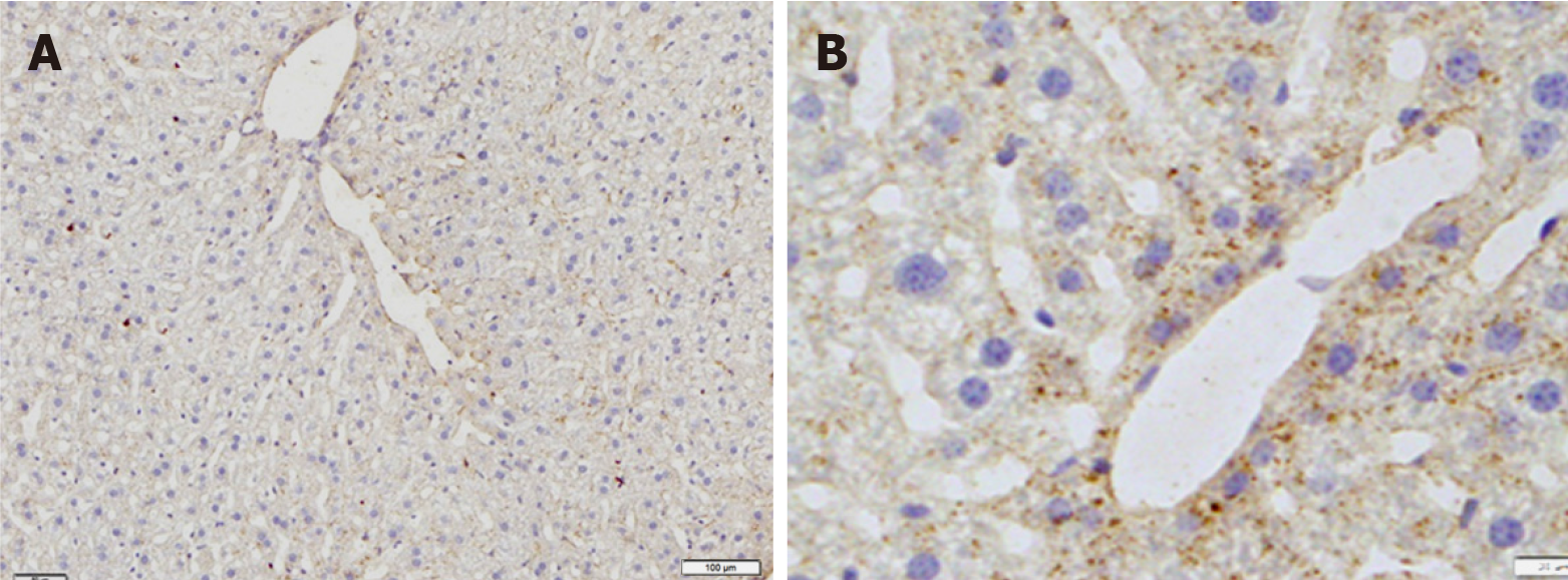
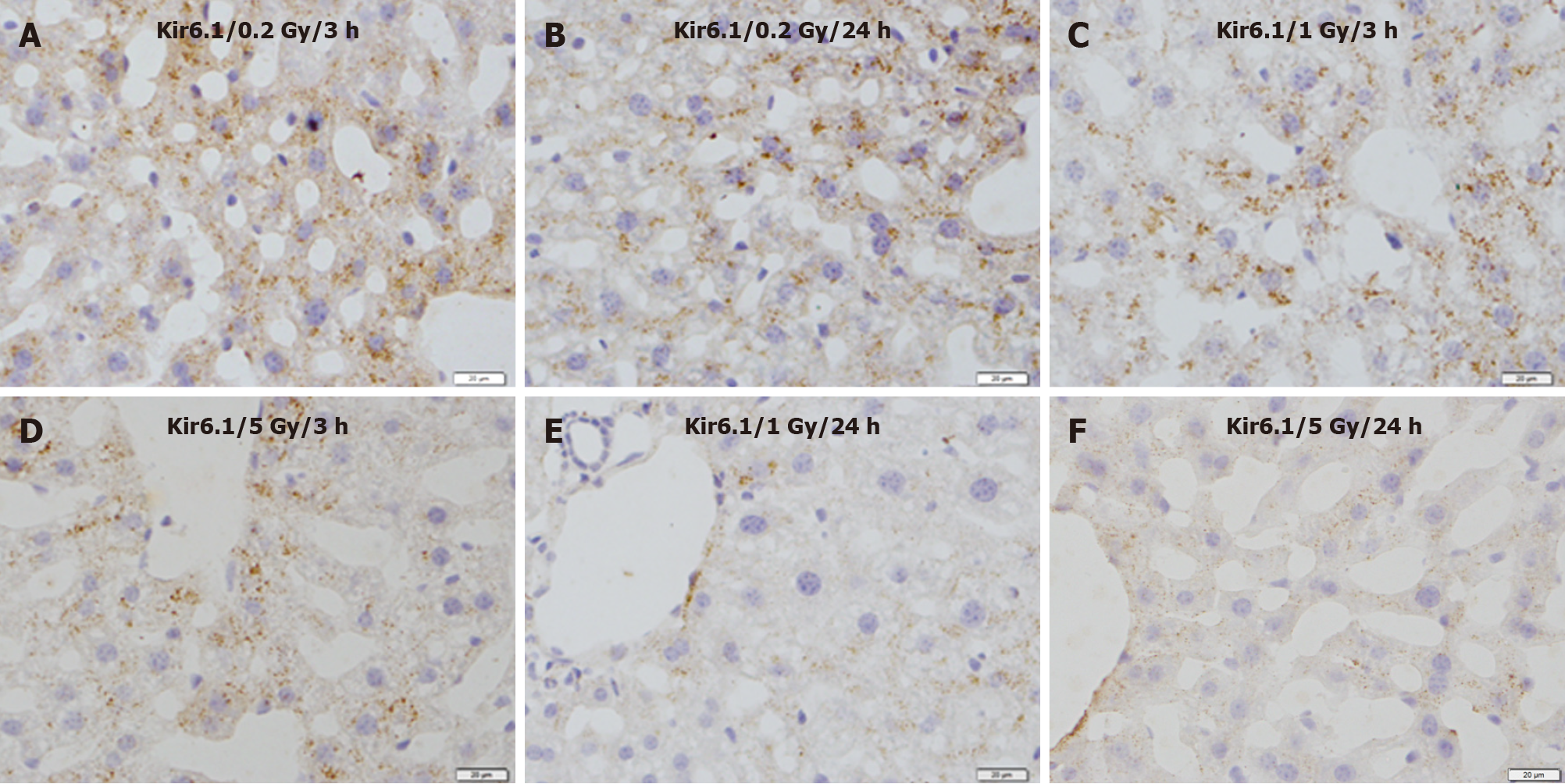
Immunohistochemical staining revealed that immunoreactivity for Kir6.1 was widely observed in hepatocytes in the control group (Figure 3A). The intensity of immunoreactivity was higher in the central vein and lower in the distal area in the cell membrane and cytoplasm of hepatocytes. Under high magnification, punctate immunoreactive products of Kir6.1 were observed in the cytoplasm of hepatocytes (Figure 3B). Compared with the control group, the expression of Kir6.1 was enhanced in the liver 3 h after exposure (Figure 4A), which almost recovered 24 h after exposure (Figure 4B) to 0.2 Gy. In the livers exposed to the medium (1 Gy) and high (5 Gy) doses of γ-ray, the expression level of Kir6.1 was decreased at 3 h (Figure 4C and D) and significantly reduced at 24 h after irradiation (Figure 4E and F).

Figure 3
Figure 3 Immunostaining for Kir6.1 in the liver of mice in the control group. A: Low magnification; B: High magnification. Bars: 100 μm (A); 20 μm (B).
Figure 4
Figure 4 Immunostaining for Kir6.1 in the liver of mice after γ-ray irradiation with different doses. A: 3 h after exposure to low dose (0.2 Gy); B: 24 h after exposure to low dose (0.2 Gy); C: 3 h after exposure to medium dose (1 Gy); D: 3 h after exposure to high dose (5 Gy); E: 24 h after exposure to medium dose (1 Gy); F: 24 h after exposure to high dose (5 Gy). Bars: 20 μm.

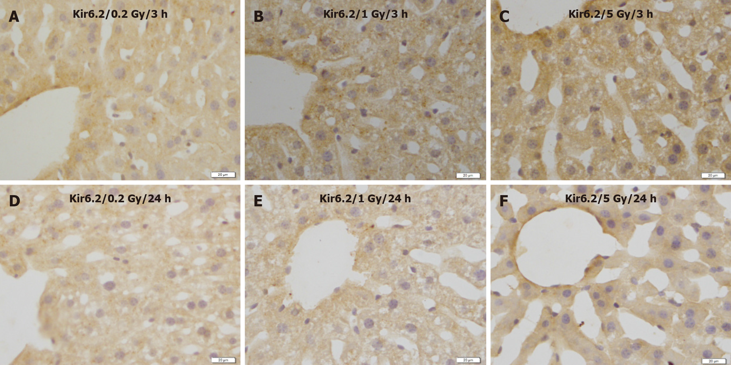
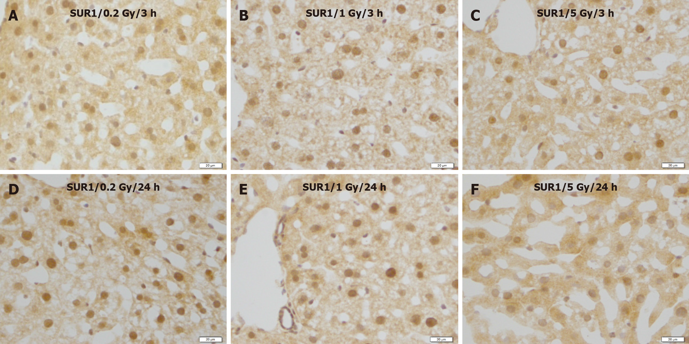
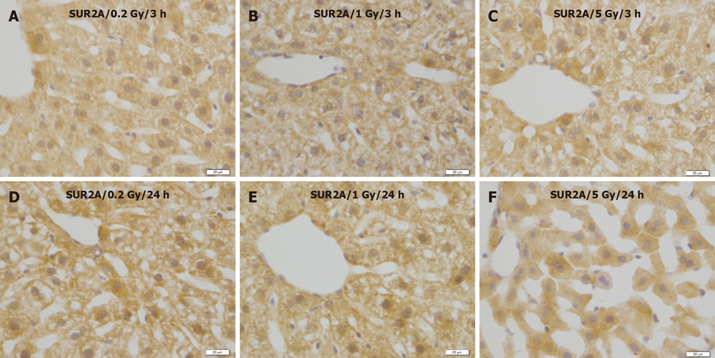
No remarkable changes were observed in the expression levels of Kir6.2 (Figure 5), SUR1 (Figure 6), or SUR2A (Figure 7) in hepatocytes at 3 and 24 h after irradiation at all doses.

Figure 5
Figure 5 Immunostaining for Kir6.2 in the liver of mice after γ-ray irradiation with different doses. Representative images show no remarkable change induced by irradiation. A: 3 h after exposure to low dose (0.2 Gy); B: 3 h after exposure to medium dose (1 Gy) ; C: 3 h after exposure to high dose (5 Gy); D: 24 h after exposure to low dose (0.2 Gy); E: 24 h after exposure to medium dose (1 Gy); F: 24 h after exposure to high dose (5 Gy). Bars: 20 μm.
Figure 6
Figure 6 Immunostaining for SUR1 in the liver of mice after γ-ray irradiation with different doses. These images show no remarkable changes in different time courses. A: 3 h after exposure to low dose (0.2 Gy); B: 3 h after exposure to medium dose (1 Gy); C: 3 h after exposure to high dose (5 Gy); D: 24 h after exposure to low dose (0.2 Gy); E: 24 h after exposure to medium dose (1 Gy); F: 24 h after exposure to high dose (5 Gy). Bars: 20 μm.
Figure 7
Figure 7 Immunostaining for SUR2A in the liver of mice after γ-ray irradiation with different doses. A: 3 h after exposure to low dose (0.2 Gy); B: 3 h after exposure to medium dose (1 Gy); C: 3 h after exposure to high dose (5 Gy); D: 24 h after exposure to low dose (0.2 Gy); E: 24 h after exposure to medium dose (1 Gy); F: 24 h after exposure to high dose (5 Gy). Bars: 20 μm.

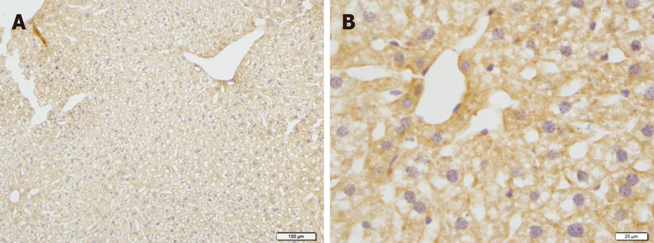
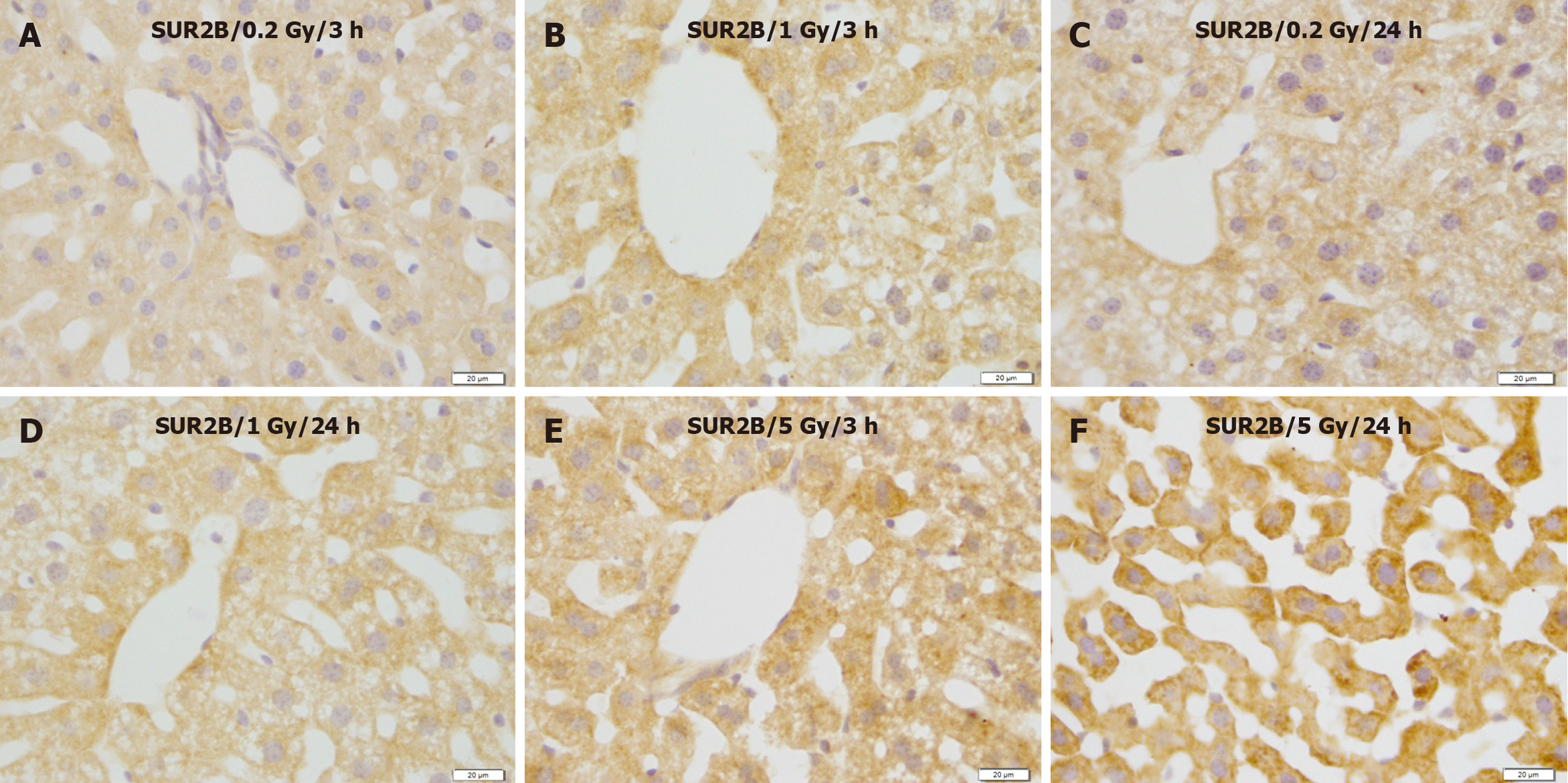
In contrast, compared with the control group (Figure 8A and B), SUR2B expression showed no apparent changes at 3 and/or 24 h after exposure to 0.2 and 1 Gy (Figure 9A-D), whereas SUR2B expression clearly increased 3 h after exposure to 5.0 Gy (Figure 9C), and more highly increased after exposure to 5.0 Gy (Figure 9E), and more highly increased 24 h after exposure to 5.0 Gy (Figure 9F).

Figure 8
Figure 8 Immunostaining for SUR2B in the liver of mice in the control groups. A: Low magnification image; B: High magnification image. Bars: 100 μm (A); 20 μm (B).
Figure 9
Figure 9 Immunostaining for SUR2B in the liver of mice after γ-ray irradiation with different doses. SUR2B expression showed no obvious change 3 and 24 h after exposure to 0.2 and 1.0 Gy, but increased remarkably 24 h after exposure to 5 Gy. A: 3 h after exposure to low dose (0.2 Gy); B: 3 h after exposure to medium dose (1 Gy); C: 24 h after exposure to low dose (0.2 Gy); D: 24 h after exposure to medium dose (1 Gy); E: 3 h after exposure to high dose (5 Gy); F: 24 h after exposure to high dose (5 Gy). Bars: 20 μm.

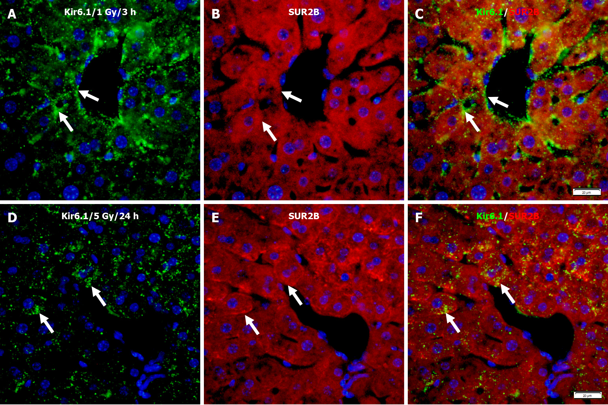
Immunofluorescence double staining for Kir6.1 and SUR2B after 1 Gy γ-ray irradiation showed only partial co-localizion of Kir6.1 with SUR2B (Figure 10A-C). However, at 24 h after mice were exposed to 5 Gy, despite a lower expression level of Kir6.1 compared to the lower dose group, almost all Kir6.1 was co-localized with SUR2B, described as punctate immunoreactivity (Figure 10D-F).

Figure 10
Figure 10  Immunofluorescence double staining for Kir6.1 and SUR2B in the liver of mice after irradiation with different doses. A-C: Representative images show the expression of Kir6.1 alone (green, A), SUR2B alone (red, B), and their co-localization (white arrows, C) 3 h after 1 Gy exposure; D-F: Representative images show the expression of Kir6.1 alone (green, D), SUR2B alone (red, E), and their co-localization (F) 24 h after 5 Gy exposure (white arrows, F). Bars: 20 μm.
DISCUSSION

Radiation effects were considered mainly due to nuclear DNA damage and their management by repair mechanisms[32]. The liver has numerous functions including bile production, ingested nutrient metabolism, waste product elimination, glycogen storage, and protein synthesis[4]. Additionally, it is radiosensitive, particularly in young animals[33]. Liver cancer, with high mortality, is one of the most common solid tumors in the world[34]. Although surgical resection is the first choice of treatment for hepatocellular carcinoma, radiation therapy is an actively used and essential treatment modality for locally advanced hepatocellular carcinoma or tumors in the upper abdomen, right lower lung, distal section of the esophagus, or whole body[4,5,35]. Liver injury induced by radiation is a significant complication of radiotherapy for liver cancer or other upper abdominal malignant tumors that have poor pharmacological therapeutic options because normal tissues exposed to radiation during radiotherapy or radioscopy will suffer injury and metabolic alterations[36]. A significant challenge in radiotherapy is to promote the tolerance of normal cells by protecting their transformation into malignant cells, thus increasing patients’ quality of life[37].

Ionizing radiation induces oxidative stress and is pivotal in the pathogenesis of cellular damage induced by radiation; dietary antioxidants were suggested to protect against irradiation-induced subsequent tissue damage[37].

Oxidative stress from generating reactive oxygen species (ROS) results in an imbalance in cells’ pro-oxidant/antioxidant status[38]. Cell survival and proliferation capacities are highly dependent on the activation and regulation through molecular components of organelles, especially mitochondria, which are pivotal in maintaining cellular homeostasis and genomic stability after irradiation[32].

Mitochondria ensure general cellular metabolism and high-energy provision via ATP and oxidative phosphorylation to maintain cell survival and homeostasis. During oxidative phosphorylation, a few electrons may react with oxygen, forming ROS and oxidative stress, thus constituting non-negligible targets for irradiation[32]. Excessive production of ROS results in mitochondria dysfunction; a series of pathological changes can be induced by radiation via direct energy deposition or reactive free radical generation[36]. Mitochondria are involved in oxidative stress-induced apoptotic cell death[39].

Upregulation of antioxidant enzymes was mimicked by treatment with the sulfonylureas tolbutamide and gliclazide (KATP channel blockers and inhibitors); the loss of KATP channel activity conferred resistance to radiation[39], suggesting a probable role for KATP channels in radiation-induced injury.

KATP channels are composed of two subunits: Kir6.x and SURs. Different combinations of these subunits are expressed in different cells and tissues and exhibit different physical characteristics[40]. KATP channels are involved in various physiological conditions, including hyperglycemia, hypoglycemia, ischemia, and hypoxia[19,40]. Kir6.1 and SUR2B are important for cellular energy and stability in the cell membrane and the mitochondrion. Mitochondria generate energy for the cell to maintaining cellular homeostasis, genomic stability, and sensitivity to irradiation[32]. Cellular oxidative stress can affect the function of potassium channels, influencing the vasomotor function in multiple disease states[41]. We have previously reported that the mitochondrial KATP channel subunits Kir6.1 and SUR2B protect heart tissues damaged by ischemia, hypoxia, or other conditions [10,11]. A recent study demonstrated that Kir6.1 and SUR2B are essential for functional brain hyperemic responses and vascular smooth muscle cell differentiation[42]. In this study, high levels of γ-ray irradiation in mice induced liver injury (Figure 2) and modulated KATP channel subunits expression, resulting in decreased Kir6.1 (Figure 4) and increased SUR2B (Figure 9) levels. These phenomena were documented using double fluorescence immunostaining (Figure 10). Although Kir6.1 had a lower expression level, the two subunits were more co-localized. High expression levels of SUR2B regulate the KATP channel in the mouse liver during high-dose irradiation. Thus, redox modulation of potassium channel activity induced by radiation is a crucial mechanism for regulating smooth muscle membrane potential, ultimately influencing hepatocyte injury.

Resent research has revealed that opening the KATP channels may increase liver tolerance to ischemia/reperfusion injury and reduce the systemic inflammatory responses[43]. The KATP channel enhances liver regeneration after partial hepatectomy[17]. However, another study showed that glibenclamide, a KATP channel blocker, prevents acute radiation-induced liver injury[44]. The effects of KATP channels in different organs and tissues are not always evident; for example, the KATP channel blocker glibenclamide ameliorated the ischemia-reperfusion injury in the rat testis, whereas 5-hydroxydecanoate does not; KATP openers mediate pharmacological post-conditioning in the heart or brain, but cannot reduce ischemia-reperfusion injury in the kidneys, intestine, and lungs[45].

Thus, the expression levels of the KATP channel subunits in the liver and their potential roles in radiation-induced liver injury still need to be investigated.

This study observed that high levels of γ-ray exposure induced liver injury in mice and modulated KATP channel subunit expression, resulting in decreased Kir6.1 and increased SUR2B levels. These findings suggest that KATP channels affect radiation-induced liver injury, and may serve as parameters for the evaluation of radiation injury. Further investigation is required to elucidate the precise roles of KATP channels in radiation-induced liver injury.

CONCLUSION

The expression of the KATP channel subunits Kir6.1 and SUR2B in the liver changed differently in response to different radiation doses, suggesting that they play a potential role during radiation-induced liver injury.

ACKNOWLEDGEMENTS

This study was partially supported by The Program of the Network-Type Joint Usage/Research Center for Radiation Disaster Medical Science of Hiroshima University, Nagasaki University. The funders played no role in the study design, data collection and analysis, decision to publish, or manuscript preparation.

Footnotes

Provenance and peer review: Unsolicited article; Externally peer reviewed.

Peer-review model: Single blind

Corresponding Author's Membership in Professional Societies: Japanese Association of Anatomists, 242307.

Specialty type: Anatomy and morphology

Country/Territory of origin: Japan

Peer-review report’s scientific quality classification

Grade A (Excellent): 0

Grade B (Very good): B

Grade C (Good): C

Grade D (Fair): 0

Grade E (Poor): 0

P-Reviewer: Emran TB, Bangladesh S-Editor: Liu JH L-Editor: Wang TQ P-Editor: Yuan YY

References
1.  Shedid SM, Abdel-Magied N, Saada HN. Role of betaine in liver injury induced by the exposure to ionizing radiation. Environ Toxicol. 2019;34:123-130.  [PubMed]  [DOI]  [Cited in This Article: ]  [Cited by in Crossref: 24]  [Cited by in F6Publishing: 24]  [Article Influence: 4.8]  [Reference Citation Analysis (0)]
2.  Kim J, Jung Y. Radiation-induced liver disease: current understanding and future perspectives. Exp Mol Med. 2017;49:e359.  [PubMed]  [DOI]  [Cited in This Article: ]  [Cited by in Crossref: 95]  [Cited by in F6Publishing: 119]  [Article Influence: 17.0]  [Reference Citation Analysis (0)]
3.  Cheng W, Xiao L, Ainiwaer A, Wang Y, Wu G, Mao R, Yang Y, Bao Y. Molecular responses of radiation-induced liver damage in rats. Mol Med Rep. 2015;11:2592-2600.  [PubMed]  [DOI]  [Cited in This Article: ]  [Cited by in Crossref: 25]  [Cited by in F6Publishing: 24]  [Article Influence: 2.7]  [Reference Citation Analysis (0)]
4.  Pan CC, Kavanagh BD, Dawson LA, Li XA, Das SK, Miften M, Ten Haken RK. Radiation-associated liver injury. Int J Radiat Oncol Biol Phys. 2010;76:S94-100.  [PubMed]  [DOI]  [Cited in This Article: ]  [Cited by in Crossref: 455]  [Cited by in F6Publishing: 481]  [Article Influence: 34.4]  [Reference Citation Analysis (0)]
5.  Chen G, Zhao Q, Yuan B, Wang B, Zhang Y, Li Z, Du S, Zeng Z. ALKBH5-Modified HMGB1-STING Activation Contributes to Radiation Induced Liver Disease via Innate Immune Response. Int J Radiat Oncol Biol Phys. 2021;111:491-501.  [PubMed]  [DOI]  [Cited in This Article: ]  [Cited by in Crossref: 7]  [Cited by in F6Publishing: 24]  [Article Influence: 8.0]  [Reference Citation Analysis (0)]
6.  Inagaki N, Tsuura Y, Namba N, Masuda K, Gonoi T, Horie M, Seino Y, Mizuta M, Seino S. Cloning and functional characterization of a novel ATP-sensitive potassium channel ubiquitously expressed in rat tissues, including pancreatic islets, pituitary, skeletal muscle, and heart. J Biol Chem. 1995;270:5691-5694.  [PubMed]  [DOI]  [Cited in This Article: ]  [Cited by in Crossref: 303]  [Cited by in F6Publishing: 322]  [Article Influence: 11.1]  [Reference Citation Analysis (0)]
7.  Ashcroft FM, Kakei M, Kelly RP. Rubidium and sodium permeability of the ATP-sensitive K+ channel in single rat pancreatic beta-cells. J Physiol. 1989;408:413-429.  [PubMed]  [DOI]  [Cited in This Article: ]  [Cited by in Crossref: 29]  [Cited by in F6Publishing: 33]  [Article Influence: 0.9]  [Reference Citation Analysis (0)]
8.  Inagaki N, Inazawa J, Seino S. cDNA sequence, gene structure, and chromosomal localization of the human ATP-sensitive potassium channel, uKATP-1, gene (KCNJ8). Genomics. 1995;30:102-104.  [PubMed]  [DOI]  [Cited in This Article: ]  [Cited by in Crossref: 46]  [Cited by in F6Publishing: 48]  [Article Influence: 1.7]  [Reference Citation Analysis (0)]
9.  Suzuki M, Kotake K, Fujikura K, Inagaki N, Suzuki T, Gonoi T, Seino S, Takata K. Kir6.1: a possible subunit of ATP-sensitive K+ channels in mitochondria. Biochem Biophys Res Commun. 1997;241:693-697.  [PubMed]  [DOI]  [Cited in This Article: ]  [Cited by in Crossref: 113]  [Cited by in F6Publishing: 117]  [Article Influence: 4.3]  [Reference Citation Analysis (0)]
10.  Zhou M, Tanaka O, Sekiguchi M, He HJ, Yasuoka Y, Itoh H, Kawahara K, Abe H. ATP-sensitive K+-channel subunits on the mitochondria and endoplasmic reticulum of rat cardiomyocytes. J Histochem Cytochem. 2005;53:1491-1500.  [PubMed]  [DOI]  [Cited in This Article: ]  [Cited by in Crossref: 30]  [Cited by in F6Publishing: 32]  [Article Influence: 1.7]  [Reference Citation Analysis (0)]
11.  Zhou M, He HJ, Suzuki R, Liu KX, Tanaka O, Sekiguchi M, Itoh H, Kawahara K, Abe H. Localization of sulfonylurea receptor subunits, SUR2A and SUR2B, in rat heart. J Histochem Cytochem. 2007;55:795-804.  [PubMed]  [DOI]  [Cited in This Article: ]  [Cited by in Crossref: 25]  [Cited by in F6Publishing: 25]  [Article Influence: 1.5]  [Reference Citation Analysis (0)]
12.  Zhou M, Tanaka O, Suzuki M, Sekiguchi M, Takata K, Kawahara K, Abe H. Localization of pore-forming subunit of the ATP-sensitive K(+)-channel, Kir6.2, in rat brain neurons and glial cells. Brain Res Mol Brain Res. 2002;101:23-32.  [PubMed]  [DOI]  [Cited in This Article: ]  [Cited by in Crossref: 61]  [Cited by in F6Publishing: 58]  [Article Influence: 2.6]  [Reference Citation Analysis (0)]
13.  Zhou M, He HJ, Tanaka O, Sekiguchi M, Kawahara K, Abe H. Localization of the ATP-sensitive K(+) channel regulatory subunits SUR2A and SUR2B in the rat brain. Neurosci Res. 2012;74:91-105.  [PubMed]  [DOI]  [Cited in This Article: ]  [Cited by in Crossref: 13]  [Cited by in F6Publishing: 15]  [Article Influence: 1.3]  [Reference Citation Analysis (0)]
14.  Zhou M, Tanaka O, Sekiguchi M, Sakabe K, Anzai M, Izumida I, Inoue T, Kawahara K, Abe H. Localization of the ATP-sensitive potassium channel subunit (Kir6. 1/uK(ATP)-1) in rat brain. Brain Res Mol Brain Res. 1999;74:15-25.  [PubMed]  [DOI]  [Cited in This Article: ]  [Cited by in Crossref: 54]  [Cited by in F6Publishing: 58]  [Article Influence: 2.3]  [Reference Citation Analysis (0)]
15.  Zhou M, He HJ, Suzuki R, Tanaka O, Sekiguchi M, Yasuoka Y, Kawahara K, Itoh H, Abe H. Expression of ATP sensitive K+ channel subunit Kir6.1 in rat kidney. Eur J Histochem. 2007;51:43-51.  [PubMed]  [DOI]  [Cited in This Article: ]
16.  Zhou M, He HJ, Tanaka O, Suzuki R, Sekiguchi M, Yasuoka Y, Kawahara K, Itoh H, Abe H. Localization of the sulphonylurea receptor subunits, SUR2A and SUR2B, in rat renal tubular epithelium. Tohoku J Exp Med. 2008;214:247-256.  [PubMed]  [DOI]  [Cited in This Article: ]  [Cited by in Crossref: 10]  [Cited by in F6Publishing: 10]  [Article Influence: 0.6]  [Reference Citation Analysis (0)]
17.  Nakagawa Y, Yoshioka M, Abe Y, Uchinami H, Ohba T, Ono K, Yamamoto Y. Enhancement of liver regeneration by adenosine triphosphate-sensitive K⁺ channel opener (diazoxide) after partial hepatectomy. Transplantation. 2012;93:1094-1100.  [PubMed]  [DOI]  [Cited in This Article: ]  [Cited by in Crossref: 13]  [Cited by in F6Publishing: 13]  [Article Influence: 1.1]  [Reference Citation Analysis (0)]
18.  Zhou M, Yoshikawa K, Akashi H, Miura M, Suzuki R, Li TS, Abe H, Bando Y. Localization of ATP-sensitive K(+) channel subunits in rat liver. World J Exp Med. 2019;9:14-31.  [PubMed]  [DOI]  [Cited in This Article: ]  [Cited by in CrossRef: 3]  [Cited by in F6Publishing: 3]  [Article Influence: 0.6]  [Reference Citation Analysis (0)]
19.  Yokota R, Tanaka M, Yamasaki K, Araki M, Miyamae M, Maeda T, Koga K, Yabuuchi Y, Sasayama S. Blockade of ATP-sensitive K+ channels attenuates preconditioning effect on myocardial metabolism in swine: myocardial metabolism and ATP-sensitive K+ channels. Int J Cardiol. 1998;67:225-236.  [PubMed]  [DOI]  [Cited in This Article: ]  [Cited by in Crossref: 7]  [Cited by in F6Publishing: 7]  [Article Influence: 0.3]  [Reference Citation Analysis (0)]
20.  Aguilar-Bryan L, Clement JP 4th, Nelson DA. Sulfonylurea receptors and ATP-sensitive potassium ion channels. Methods Enzymol. 1998;292:732-744.  [PubMed]  [DOI]  [Cited in This Article: ]  [Cited by in Crossref: 12]  [Cited by in F6Publishing: 11]  [Article Influence: 0.5]  [Reference Citation Analysis (0)]
21.  Babenko AP, Gonzalez GC, Bryan J. Hetero-concatemeric KIR6.X4/SUR14 channels display distinct conductivities but uniform ATP inhibition. J Biol Chem. 2000;275:31563-31566.  [PubMed]  [DOI]  [Cited in This Article: ]  [Cited by in Crossref: 21]  [Cited by in F6Publishing: 22]  [Article Influence: 0.9]  [Reference Citation Analysis (0)]
22.  Shyng S, Nichols CG. Octameric stoichiometry of the KATP channel complex. J Gen Physiol. 1997;110:655-664.  [PubMed]  [DOI]  [Cited in This Article: ]  [Cited by in Crossref: 391]  [Cited by in F6Publishing: 360]  [Article Influence: 13.3]  [Reference Citation Analysis (0)]
23.  Seino S. Physiology and pathophysiology of K(ATP) channels in the pancreas and cardiovascular system: a review. J Diabetes Complications. 2003;17:2-5.  [PubMed]  [DOI]  [Cited in This Article: ]  [Cited by in Crossref: 26]  [Cited by in F6Publishing: 26]  [Article Influence: 1.2]  [Reference Citation Analysis (0)]
24.  Tricarico D, Mele A, Lundquist AL, Desai RR, George AL Jr, Conte Camerino D. Hybrid assemblies of ATP-sensitive K+ channels determine their muscle-type-dependent biophysical and pharmacological properties. Proc Natl Acad Sci U S A. 2006;103:1118-1123.  [PubMed]  [DOI]  [Cited in This Article: ]  [Cited by in Crossref: 49]  [Cited by in F6Publishing: 53]  [Article Influence: 2.9]  [Reference Citation Analysis (0)]
25.  Pocai A, Lam TK, Gutierrez-Juarez R, Obici S, Schwartz GJ, Bryan J, Aguilar-Bryan L, Rossetti L. Hypothalamic K(ATP) channels control hepatic glucose production. Nature. 2005;434:1026-1031.  [PubMed]  [DOI]  [Cited in This Article: ]  [Cited by in Crossref: 472]  [Cited by in F6Publishing: 473]  [Article Influence: 24.9]  [Reference Citation Analysis (0)]
26.  Malhi H, Irani AN, Rajvanshi P, Suadicani SO, Spray DC, McDonald TV, Gupta S. KATP channels regulate mitogenically induced proliferation in primary rat hepatocytes and human liver cell lines. Implications for liver growth control and potential therapeutic targeting. J Biol Chem. 2000;275:26050-26057.  [PubMed]  [DOI]  [Cited in This Article: ]  [Cited by in Crossref: 68]  [Cited by in F6Publishing: 73]  [Article Influence: 3.0]  [Reference Citation Analysis (0)]
27.  Luo L, Yan C, Urata Y, Hasan AS, Goto S, Guo CY, Zhang S, Li TS. Dose-dependency and reversibility of radiation-induced injury in cardiac explant-derived cells of mice. Sci Rep. 2017;7:40959.  [PubMed]  [DOI]  [Cited in This Article: ]  [Cited by in Crossref: 3]  [Cited by in F6Publishing: 3]  [Article Influence: 0.4]  [Reference Citation Analysis (0)]
28.  Mezaki Y, Yamaguchi N, Yoshikawa K, Miura M, Imai K, Itoh H, Senoo H. Insoluble, speckled cytosolic distribution of retinoic acid receptor alpha protein as a marker of hepatic stellate cell activation in vitro. J Histochem Cytochem. 2009;57:687-699.  [PubMed]  [DOI]  [Cited in This Article: ]  [Cited by in Crossref: 19]  [Cited by in F6Publishing: 20]  [Article Influence: 1.3]  [Reference Citation Analysis (0)]
29.  Troeger JS, Mederacke I, Gwak GY, Dapito DH, Mu X, Hsu CC, Pradere JP, Friedman RA, Schwabe RF. Deactivation of hepatic stellate cells during liver fibrosis resolution in mice. Gastroenterology. 2012;143:1073-83.e22.  [PubMed]  [DOI]  [Cited in This Article: ]  [Cited by in Crossref: 380]  [Cited by in F6Publishing: 347]  [Article Influence: 28.9]  [Reference Citation Analysis (0)]
30.  Stewart RK, Dangi A, Huang C, Murase N, Kimura S, Stolz DB, Wilson GC, Lentsch AB, Gandhi CR. A novel mouse model of depletion of stellate cells clarifies their role in ischemia/reperfusion- and endotoxin-induced acute liver injury. J Hepatol. 2014;60:298-305.  [PubMed]  [DOI]  [Cited in This Article: ]  [Cited by in Crossref: 73]  [Cited by in F6Publishing: 77]  [Article Influence: 7.7]  [Reference Citation Analysis (0)]
31.  Kavanagh JN, Waring EJ, Prise KM. Radiation responses of stem cells: targeted and non-targeted effects. Radiat Prot Dosimetry. 2015;166:110-117.  [PubMed]  [DOI]  [Cited in This Article: ]  [Cited by in Crossref: 2]  [Cited by in F6Publishing: 2]  [Article Influence: 0.2]  [Reference Citation Analysis (0)]
32.  Averbeck D, Rodriguez-Lafrasse C. Role of Mitochondria in Radiation Responses: Epigenetic, Metabolic, and Signaling Impacts. Int J Mol Sci. 2021;22.  [PubMed]  [DOI]  [Cited in This Article: ]  [Cited by in Crossref: 17]  [Cited by in F6Publishing: 69]  [Article Influence: 23.0]  [Reference Citation Analysis (0)]
33.  Zaher NH, Salem AA, Ismail AF. Novel amino acid derivatives bearing thieno[2,3-d]pyrimidine moiety down regulate NF-κB in γ-irradiation mediated rat liver injury. J Photochem Photobiol B. 2016;165:328-339.  [PubMed]  [DOI]  [Cited in This Article: ]  [Cited by in Crossref: 10]  [Cited by in F6Publishing: 10]  [Article Influence: 1.3]  [Reference Citation Analysis (0)]
34.  Yang W, Shao L, Zhu S, Li H, Zhang X, Ding C, Wu X, Xu R, Yue M, Tang J, Kuang B, Fan G, Zhu Q, Zeng H. Transient Inhibition of mTORC1 Signaling Ameliorates Irradiation-Induced Liver Damage. Front Physiol. 2019;10:228.  [PubMed]  [DOI]  [Cited in This Article: ]  [Cited by in Crossref: 3]  [Cited by in F6Publishing: 4]  [Article Influence: 0.8]  [Reference Citation Analysis (0)]
35.  Doi H, Masai N, Uemoto K, Suzuki O, Shiomi H, Tatsumi D, Oh RJ. Validation of the liver mean dose in terms of the biological effective dose for the prevention of radiation-induced liver damage. Rep Pract Oncol Radiother. 2017;22:303-309.  [PubMed]  [DOI]  [Cited in This Article: ]  [Cited by in Crossref: 6]  [Cited by in F6Publishing: 3]  [Article Influence: 0.4]  [Reference Citation Analysis (0)]
36.  Zhu W, Zhang X, Yu M, Lin B, Yu C. Radiation-induced liver injury and hepatocyte senescence. Cell Death Discov. 2021;7:244.  [PubMed]  [DOI]  [Cited in This Article: ]  [Cited by in Crossref: 4]  [Cited by in F6Publishing: 23]  [Article Influence: 7.7]  [Reference Citation Analysis (0)]
37.  Pradeep K, Park SH, Ko KC. Hesperidin a flavanoglycone protects against gamma-irradiation induced hepatocellular damage and oxidative stress in Sprague-Dawley rats. Eur J Pharmacol. 2008;587:273-280.  [PubMed]  [DOI]  [Cited in This Article: ]  [Cited by in Crossref: 69]  [Cited by in F6Publishing: 70]  [Article Influence: 4.4]  [Reference Citation Analysis (0)]
38.  Bhosle SM, Huilgol NG, Mishra KP. Enhancement of radiation-induced oxidative stress and cytotoxicity in tumor cells by ellagic acid. Clin Chim Acta. 2005;359:89-100.  [PubMed]  [DOI]  [Cited in This Article: ]  [Cited by in Crossref: 83]  [Cited by in F6Publishing: 70]  [Article Influence: 3.7]  [Reference Citation Analysis (0)]
39.  Gier B, Krippeit-Drews P, Sheiko T, Aguilar-Bryan L, Bryan J, Düfer M, Drews G. Suppression of KATP channel activity protects murine pancreatic beta cells against oxidative stress. J Clin Invest. 2009;119:3246-3256.  [PubMed]  [DOI]  [Cited in This Article: ]  [Cited by in Crossref: 13]  [Cited by in F6Publishing: 37]  [Article Influence: 2.5]  [Reference Citation Analysis (0)]
40.  Seino S, Miki T. Physiological and pathophysiological roles of ATP-sensitive K+ channels. Prog Biophys Mol Biol. 2003;81:133-176.  [PubMed]  [DOI]  [Cited in This Article: ]  [Cited by in Crossref: 371]  [Cited by in F6Publishing: 374]  [Article Influence: 17.8]  [Reference Citation Analysis (0)]
41.  Liu Y, Gutterman DD. Oxidative stress and potassium channel function. Clin Exp Pharmacol Physiol. 2002;29:305-311.  [PubMed]  [DOI]  [Cited in This Article: ]  [Cited by in Crossref: 118]  [Cited by in F6Publishing: 119]  [Article Influence: 5.4]  [Reference Citation Analysis (0)]
42.  Ando K, Tong L, Peng D, Vázquez-Liébanas E, Chiyoda H, He L, Liu J, Kawakami K, Mochizuki N, Fukuhara S, Grutzendler J, Betsholtz C. KCNJ8/ABCC9-containing K-ATP channel modulates brain vascular smooth muscle development and neurovascular coupling. Dev Cell. 2022;57:1383-1399.e7.  [PubMed]  [DOI]  [Cited in This Article: ]  [Cited by in Crossref: 2]  [Cited by in F6Publishing: 18]  [Article Influence: 9.0]  [Reference Citation Analysis (0)]
43.  Nogueira MA, Coelho AM, Sampietre SN, Patzina RA, Pinheiro da Silva F, D'Albuquerque LA, Machado MC. Beneficial effects of adenosine triphosphate-sensitive K+ channel opener on liver ischemia/reperfusion injury. World J Gastroenterol. 2014;20:15319-15326.  [PubMed]  [DOI]  [Cited in This Article: ]  [Cited by in CrossRef: 15]  [Cited by in F6Publishing: 15]  [Article Influence: 1.5]  [Reference Citation Analysis (0)]
44.  Liu H, Wang S, Wu Z, Huang Z, Chen WY, Yang Y, Cui J, Liu C, Zhao H, Guo J, Zhang P, Gao F, Li B, Cai J. Glibenclamide, a diabetic drug, prevents acute radiation induced liver injury of mice via up-regulating intracellular ROS and subsequently activating Akt-NF-κB pathway. Oncotarget. 2017;8:40568-40582.  [PubMed]  [DOI]  [Cited in This Article: ]  [Cited by in Crossref: 14]  [Cited by in F6Publishing: 11]  [Article Influence: 1.6]  [Reference Citation Analysis (0)]
45.  Shimizu S, Oikawa R, Tsounapi P, Inoue K, Shimizu T, Tanaka K, Martin DT, Honda M, Sejima T, Tomita S, Saito M. Blocking of the ATP sensitive potassium channel ameliorates the ischaemia-reperfusion injury in the rat testis. Andrology. 2014;2:458-465.  [PubMed]  [DOI]  [Cited in This Article: ]  [Cited by in Crossref: 18]  [Cited by in F6Publishing: 18]  [Article Influence: 1.8]  [Reference Citation Analysis (0)]