Minireviews Open Access
Copyright ©The Author(s) 2024. Published by Baishideng Publishing Group Inc. All rights reserved.
World J Exp Med. Jun 20, 2024; 14(2): 88867
Published online Jun 20, 2024. doi: 10.5493/wjem.v14.i2.88867
Ovarian function in patients with systemic lupus erythematosus: Pathogenesis, drug application and prospective therapies
Min Xu, Li-Li Tian, Xiao-Liu Li, Hong-Wei Chen, Department of Rheumatology and Immunology, Nanjing Drum Tower Hospital, Clinical College of Nanjing University of Chinese Medicine, Nanjing 210008, Jiangsu Province, China
Cheng Bao, School of Life Science, Nanjing Normal University, Nanjing 210023, Jiangsu Province, China
Cheng Bao, Department of Rheumatology and Immunology, Nanjing Drum Tower Hospital, Nanjing 210008, Jiangsu Province, China
Hai-Wei Zhang, Department of Rheumatology and Immunology, Nanjing Pukou People’s Hospital, Nanjing 211800, Jiangsu Province, China
Hong-Wei Chen, Department of Rheumatology and Immunology, Nanjing Drum Tower Hospital, The Affiliated Hospital of School of Medicine, Nanjing University, Nanjing 210008, Jiangsu Province, China
ORCID number: Hong-Wei Chen (0000-0002-9400-6053).
Author contributions: Xu M, Tian LL, Li XL, and Bao C wrote the manuscript; Zhang HW performed the research; Chen HW designed the review and were responsible for the final proofreading; All authors have read and approve the final manuscript.
Supported by the National Natural Science Foundation of China, No. 82271843; and Key Project supported by Medical Science and Technology Development Foundation, Nanjing Department of Health, No. ZKX20019.
Conflict-of-interest statement: All authors declare no conflicts of interest for this article.
Open-Access: This article is an open-access article that was selected by an in-house editor and fully peer-reviewed by external reviewers. It is distributed in accordance with the Creative Commons Attribution NonCommercial (CC BY-NC 4.0) license, which permits others to distribute, remix, adapt, build upon this work non-commercially, and license their derivative works on different terms, provided the original work is properly cited and the use is non-commercial. See: https://creativecommons.org/Licenses/by-nc/4.0/
Corresponding author: Hong-Wei Chen, PhD, Associate Professor, Department of Rheumatology and Immunology, Nanjing Drum Tower Hospital, The Affiliated Hospital of School of Medicine, Nanjing University, No. 321 Zhongshan Road, Nanjing 210008, Jiangsu Province, China. chenhw@nju.edu.cn
Received: October 12, 2023
Revised: February 27, 2024
Accepted: April 9, 2024
Published online: June 20, 2024
Processing time: 251 Days and 3.7 Hours

Abstract

Systemic lupus erythematosus (SLE) is a chronic autoimmune disease in which multiple organs are damaged that prevails in fertile women. Currently, glucocorticoids and immunosuppressants are widely used to treat SLE patients. However, ovarian dysfunction occurs following the use of these drugs in women with SLE. Here, we summarize recent progress in terms of understanding ovarian injury, the effects of drug application and strategies to improve ovarian function in women with SLE. This review could be helpful to precisely cure SLE in women desiring to have offspring.

Key Words: Systemic lupus erythematosus; Ovarian reserve; Ovarian insufficiency; Mesenchymal stem cells; Fertility; Autoimmune disease

Core Tip: Systemic lupus erythematosus (SLE) is an autoimmune disease that often occurs in women of childbearing age. Disorders of the immune system and clinical treatment drugs can affect the ovarian and reproductive functions of female patients. Herein, we summarize the research progress on SLE combined with ovarian dysfunction, hoping to provide a reference for the clinical treatment of patients with SLE for ovarian function and fertility needs.


INTRODUCTION

Systemic lupus erythematosus (SLE) is an autoimmune disease characterized by autoreactive activity of the immune system, the production of a variety of autoantibodies, abundant deposition of immune complexes, and damage to multiple tissues and organs. The ovaries, as female gonads, are the organs from which estrogen is secreted. Follicles mature and ovulate, playing an important role in endocrine and reproductive functions. SLE commonly occurs in women between 20 and 40 years of age. If immune complexes are deposited in ovaries, they can cause ovarian damage, manifesting as low ovarian reserve[1]. Currently, drugs such as tacrolimus, mycophenolate mofetil, cyclophosphamide, and prednisolone are the main treatment options for patients with SLE. However, some of these drugs, such as cyclophosphamide, have ovarian toxicity. They may cause less menstruation, amenorrhea, infertility, low ovarian reserve, premature ovarian failure and other clinical manifestations in women with SLE. Therefore, this review briefly summarizes the abnormal changes in ovarian function indicators, the ovarian damage caused by clinical medications, and the measures to improve ovarian function in female patients with SLE in recent years, hoping to provide insights into the ovarian reserve function of patients with SLE and the clinical treatment for patients with SLE with reproductive needs.

ABNORMAL OVARIAN FUNCTION IN PATIENTS WITH SLE

Ovarian reserve refers to the quantity and quality of oocytes in the ovary and is closely related to female fertility[2]. Clinically, the most commonly used indicators to evaluate ovarian reserve include the menstrual cycle, antral follicle count (AFC), ovarian volume (OV), follicle stimulating hormone (FSH), corpus luteum luteinizing hormone (LH) and anti-Müllerian hormone (AMH).

Menstrual cycle

A woman’s menstrual cycle can reflect her ovarian function and reproductive capacity. The length of the menstrual cycle, regular or disordered cycle, the amount of menstrual flow, and amenorrhea can all reflect ovarian function and hormone secretion levels. Female patients with SLE have been suggested to be more likely to suffer from menstrual disorders and even persistent amenorrhea. In a recent study of 40 SLE patients, 45% of the patients (18 patients) were reported to have irregular menstruation, including 11 patients with oligomenorrhea, 1 patient with irregular menstruation, 4 patients with oligomenorrhea and irregular menstruation, and 1 patient with amenorrhea, and the rate of menstrual abnormalities was significantly greater in these patients than in healthy control individuals[3]. This is consistent with the studies carried out by Shabanova[4] and Girbash[5]. In addition, the systemic lupus erythematosus disease activity index (SLEDAI) score of SLE patients with irregular menstrual cycles was notably greater than that of SLE patients with regular menstrual cycles.

Ovarian morphology

The ovarian morphology can indicate its reserve function. The number of primordial follicles in the ovary represents a woman’s ovarian reserve, and the AFC is generally used to quantify ovarian function[6]. A decrease in the number of AFCs often indicates that the responsiveness and reserve function of the ovary are reduced[6]. In addition, a smaller OV will cause lower sex hormone levels and ovarian responsiveness. Therefore, the OV is also an important indicator of ovarian reserve[7]. Girbash et al[5] compared 50 SLE patients with 50 healthy control individuals of similar age and found that the AFCs and OVs of SLE patients were markedly reduced, and both were related to age.

Sex hormones

Follicles are mainly composed of granulosa cells and eggs. Granulosa cells play a vital role in egg development, differentiation and maturation. Estradiol (E2) is secreted by follicular granulosa cells and can be used as an indicator of ovarian reserve. However, this indicator is susceptible to ovarian diseases and other indicators, and thus, it cannot be simply evaluated alone. Since the ovaries are regulated by the hypothalamic-pituitary-gonadal (HPG) axis, when the ovarian reserve is reduced, negative feedback occurs to promote FSH and LH secretion. Therefore, in clinical practice, FSH, FSH/LH, and E2 are often jointly tested as biochemical indicators for evaluating ovarian function[7]. If a patient’s ovarian reserve decreases, her E2 levels decrease, and her FSH and FSH/LH values increase accordingly. This phenomenon corresponds to the results after ovarian damage in SLE patients. In a prospective study, researchers measured FSH, E2, and LH on the third day of the menstrual cycle. They found that E2 levels were reduced in SLE patients, and FSH and LH levels were significantly greater than those in healthy control individuals[8].

Anti-Müllerian hormone

AMH is also secreted by follicular granulosa cells and remains relatively stable throughout the menstrual cycle. It is more sensitive and specific than other biochemical indicators and is therefore widely used as a direct indicator of ovarian reserve. Lawrenz et al[9] measured the AMH values of 33 premenopausal SLE patients and compared them with the AMH values of 33 age-matched healthy people. The authors found that the AMH levels in the SLE group were notably lower than those in the healthy group. Furthermore, they found that there was no correlation between the AMH value and the SLEDAI score. Conversely, in another case-control study of 40 SLE patients of childbearing age, the AMH level was significantly lower in patients with SLE than in healthy control individuals and was negatively correlated with SLEDAI[3]. Thus, the correlation between AMH levels and disease activity in SLE patients still needs further large-scale testing to draw an accurate conclusion.

EFFECTS OF SLE MEDICATION ON OVARIAN FUNCTION
Glucocorticoids

Glucocorticoids (GCs) are the preferred medication for treating SLE patients and can quickly suppress immune responses and reduce the damage of inflammatory reactions to the body, thereby rapidly alleviating the condition of SLE. However, GCs (especially high-dose GCs) can decrease levels of ovarian-related sex hormones and AMH secretion, decrease AFC and OV, and cause amenorrhea, and thus, GCs can impair ovarian function in patients with SLE. All these changes may be related to the inhibition of the HPG axis function by GC[10].

Whirledge et al[11] assumed that GCs affect ovarian function through the following three mechanisms. GCs influence ovarian function indirectly through altering the levels of circulating gonadotropins by acting on the hypothalamus and pituitary. Excessive GC levels inhibit gonadotropin-releasing hormone secretion, leading to hypogonadotropic hypogonadism. The second mechanism of glucocorticoid regulation is also indirect and affects the levels of metabolic hormones and growth factors, such as insulin-like growth factor-1. Finally, GCs regulate ovarian function by binding to GC receptors on ovarian cells, affecting LH activity and steroid biosynthesis. It can be concluded that GCs can extensively act on all links of the HPO axis to cause damage to ovarian function.

Cyclophosphamide

Cyclophosphamide (CTX), a commonly used immunosuppressant, unfortunately has strong gonadal toxicity and can lead to amenorrhea and infertility[12]. Boumpas et al[13] reported that 28% of SLE patients had persistent amenorrhea after CTX treatment, and 8% of the patients had temporary amenorrhea. SLE patients receiving high-dose CTX treatment were more likely to have persistent amenorrhea than were those receiving low-dose CTX treatment (39% vs 12%, respectively). In addition, amenorrhea occurs in 12% of patients younger than 25 years and in 62% of patients older than 31 years. The above studies have shown that the ovarian damage caused by CTX is closely related to the age of SLE patients at the start of CTX treatment and the cumulative dose over treatment time, which has been corroborated by the results of others[14,15]. Therefore, CTX should be used with caution in SLE patients of childbearing age who are willing to have children.

CTX can activate the PI3K-AKT-mTOR signaling pathway and increase the phosphorylation of AKT and other related proteins. Thus, it can directly affect the oocytes and granulosa cells of primordial follicles and accelerate the activation of primordial follicles[16]. Moreover, CTX-induced ovarian toxicity may be mediated by an inflammatory response, which can be caused by increasing the levels of proinflammatory cytokines [interleukin (IL)-6, IL-8, TNFα, etc.] and decreasing the levels of anti-inflammatory factors such as IL-10[17]. In addition, CTX changes the expression of pro- and antiapoptotic genes, leading to increased apoptosis in the ovary[18]. In conclusion, CTX induces oocyte damage and apoptosis in granulosa cells and follicles, resulting in premature depletion of follicular reserves.

In addition, other drugs used to treat SLE, such as hydroxychloroquine, tripterygium glycosides, mycophenolate mofetil and tacrolimus, are rarely reported to have effects on the ovary and need to be further studied.

MEASURES TO IMPROVE OVARIAN FUNCTION IN PATIENTS WITH SLE

The recurrent attacks of SLE can significantly exacerbate the damage to affected tissues and organs. In addition, the ovarian damage resulting from the long-term application of glucocorticoids and immunosuppressants cannot be ignored. For patients with SLE of childbearing age, it is essential to protect their ovarian function and maintain fertility, thereby necessitating the urgent development of a treatment strategy to improve ovarian damage.

The clinical application of CTX enhances the recruitment of follicles, eventually leading to ovarian dysfunction. Therefore, inhibiting ovulation reduces the ovarian toxicity of CTX during treatment. Gonadotropin releasing hormone (GnRH) is secreted from the hypothalamus to the pituitary portal circulation, where it stimulates the pituitary to secrete FSH and LH and maintain the normal menstrual cycle. GnRH analog (GnRH-a) is a synthetic agonist of GnRH that can competitively bind to the gonadotropin-releasing hormone receptor on the pituitary gland and inhibit the secretion of FSH and LH, thus inhibiting ovulation[19]. In addition, clinical studies have reported that GnRH-a combined with CTX in the treatment of SLE reduces ovarian exposure to alkylation reagents, resulting in a reduction in ovarian damage[20,21].

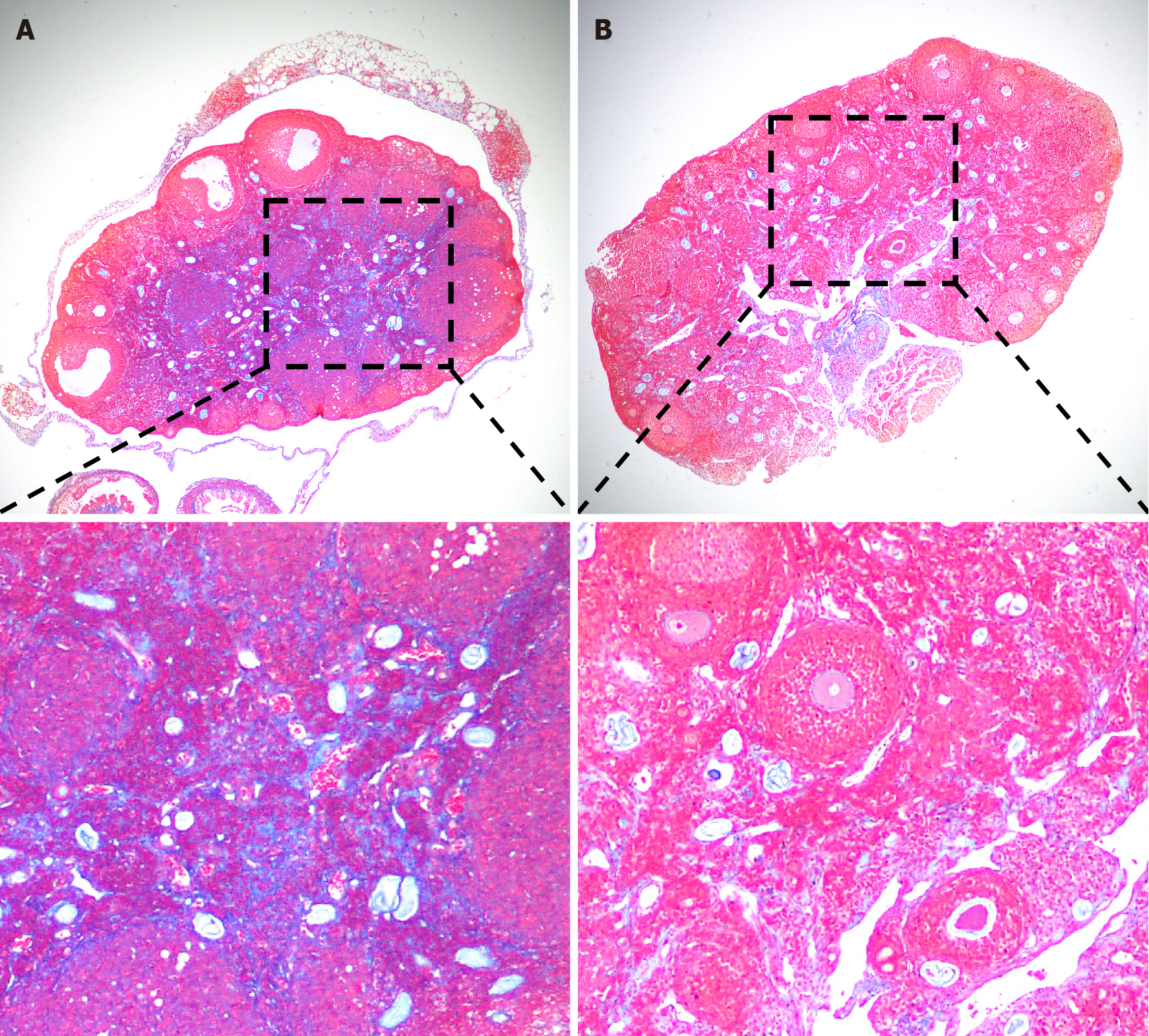
Severe ovarian damage profoundly impacts the entire reproductive system and can even result in infertility in severe cases. Currently, the cryopreservation of embryos and oocytes is considered a promising approach for preserving fertility. However, the social, ethical, and technical challenges associated with this strategy cannot be neglected. Mesenchymal stem cells (MSCs) are multipotent adult stem cells with low immunogenicity, self-renewal to maintain their stem cell properties, and multidirectional differentiation ability[22]. MSCs can migrate into the damaged ovary to increase the production of bioactive factors[23,24], such as vascular endothelial growth factor, hepatocyte growth factor, insulin-like growth factor, and other growth factors that promote immunomodulation, anti-inflammation, angiogenesis, anti-apoptosis, and anti-fibrosis. In recent years, MSC therapy has gradually been applied to treat ovarian insufficiency[25-27], and its efficacy has been confirmed by clinical trials. Using a polycystic ovary syndrome (PCOS) mouse model, Chugh et al[27] confirmed that intraovarian injection of bone marrow mesenchymal cells significantly reduced steroid gene expression, thereby inhibiting the inflammatory response and restoring ovarian function. Transplantation of human umbilical cord MSCs (UC-MSCs) into patients with ovarian insufficiency can increase E2 levels and the number of follicles, improve follicular development, and result in successful clinical pregnancy[27]. Furthermore, our recent experiments demonstrated that UC-MSC transplantation improves ovarian function by inhibiting ovarian fibrosis in SLE model mice (Figure 1). Hence, MSCs are of great significance in the treatment of immune-related ovarian insufficiency, such as pregnancy complicated with SLE. Clinical data from trials carrying out MSC therapy to treat women with SLE are expected to validate this new approach in the future.

Figure 1
Figure 1 Umbilical cord mesenchymal stem cell (UC-MSC) transplantation reduced ovarian fibrosis in MRL/Lpr (MRL/Mpj-Faslpr/J, #000485, The Jackon Laboratories, United States) mice. A: Representative masson staining of ovaries from MRL/Lpr mice in the PBS group; B: Representative masson staining of ovaries from MRL/Lpr mice in the umbilical cord mesenchymal stem cell transplantation group. Magnification: 40 × (top) and 100 × (bottom).
CONCLUSION

In summary, SLE damages ovarian and reproductive function in female patients of childbearing age. Disorders of the immune system can lead to autoimmune oophoritis, which injures the ovaries and reduces the ovarian reserve. Additionally, when treated with glucocorticoids, cyclophosphamide, triptolide polyglycosides, etc., gonadal toxicity will occur, resulting in HPO axis disorders and even adverse pregnancy outcomes. To better address ovarian dysfunction in SLE patients, the key unanswered question is how proinflammatory niches cause ovarian damage and loss of function. Therefore, it is important to seek safe and effective treatments for SLE and ovarian insufficiency. MSCs have been widely used to treat SLE and have great potential for treating ovarian insufficiency-related diseases. They are expected to become an ideal and reliable treatment for SLE combined with ovarian insufficiency in the near future.

Footnotes

Provenance and peer review: Invited article; Externally peer reviewed.

Peer-review model: Single blind

Specialty type: Immunology

Country/Territory of origin: China

Peer-review report’s classification

Scientific Quality: Grade B

Novelty: Grade B

Creativity or Innovation: Grade B

Scientific Significance: Grade B

P-Reviewer: Dauyey K, Kazakhstan S-Editor: Liu JH L-Editor: A P-Editor: Yuan YY

References
1.  Kiriakidou M, Ching CL. Systemic Lupus Erythematosus. Ann Intern Med. 2020;172:ITC81-ITC96.  [PubMed]  [DOI]  [Cited in This Article: ]  [Cited by in Crossref: 176]  [Cited by in F6Publishing: 385]  [Article Influence: 96.3]  [Reference Citation Analysis (0)]
2.  Findlay JK, Hutt KJ, Hickey M, Anderson RA. What is the "ovarian reserve"? Fertil Steril. 2015;103:628-630.  [PubMed]  [DOI]  [Cited in This Article: ]  [Cited by in Crossref: 38]  [Cited by in F6Publishing: 31]  [Article Influence: 3.4]  [Reference Citation Analysis (0)]
3.  Gao H, Ma J, Wang X, Lv T, Liu J, Ren Y, Li Y, Zhang Y. Preliminary study on the changes of ovarian reserve, menstruation, and lymphocyte subpopulation in systemic lupus erythematosus (SLE) patients of childbearing age. Lupus. 2018;27:445-453.  [PubMed]  [DOI]  [Cited in This Article: ]  [Cited by in Crossref: 11]  [Cited by in F6Publishing: 13]  [Article Influence: 1.9]  [Reference Citation Analysis (0)]
4.  Shabanova SS, Ananieva LP, Alekberova ZS, Guzov II. Ovarian function and disease activity in patients with systemic lupus erythematosus. Clin Exp Rheumatol. 2008;26:436-441.  [PubMed]  [DOI]  [Cited in This Article: ]
5.  Girbash EF, Abdelwahab SM, Fahmi DS, Abdeldayem HM, Ghonaim R, Atta DS. Preliminary study on anti-Müllerian hormone, antral follicle count, menstruation and lymphocyte subsets in systemic lupus erythematosus patients. Int J Gynaecol Obstet. 2022;159:129-135.  [PubMed]  [DOI]  [Cited in This Article: ]  [Reference Citation Analysis (0)]
6.  Campbell S. Ultrasound Evaluation in Female Infertility: Part 1, the Ovary and the Follicle. Obstet Gynecol Clin North Am. 2019;46:683-696.  [PubMed]  [DOI]  [Cited in This Article: ]  [Cited by in Crossref: 6]  [Cited by in F6Publishing: 6]  [Article Influence: 1.2]  [Reference Citation Analysis (0)]
7.  Practice Committee of the American Society for Reproductive Medicine. Practice Committee of the American Society for Reproductive Medicine. Testing and interpreting measures of ovarian reserve: a committee opinion. Fertil Steril. 2020;114:1151-1157.  [PubMed]  [DOI]  [Cited in This Article: ]  [Cited by in Crossref: 42]  [Cited by in F6Publishing: 50]  [Article Influence: 12.5]  [Reference Citation Analysis (0)]
8.  Ulug P, Oner G, Kasap B, Akbas EM, Ozcicek F. Evaluation of ovarian reserve tests in women with systemic lupus erythematosus. Am J Reprod Immunol. 2014;72:85-88.  [PubMed]  [DOI]  [Cited in This Article: ]  [Cited by in Crossref: 12]  [Cited by in F6Publishing: 14]  [Article Influence: 1.4]  [Reference Citation Analysis (0)]
9.  Lawrenz B, Henes J, Henes M, Neunhoeffer E, Schmalzing M, Fehm T, Kïtter I. Impact of systemic lupus erythematosus on ovarian reserve in premenopausal women: evaluation by using anti-Muellerian hormone. Lupus. 2011;20:1193-1197.  [PubMed]  [DOI]  [Cited in This Article: ]  [Cited by in Crossref: 94]  [Cited by in F6Publishing: 98]  [Article Influence: 7.5]  [Reference Citation Analysis (0)]
10.  Lourenço DMR, Araújo DB, Aikawa NE, Yamakami LYS, Borba EF, Maciel GAR, Soares-Junior JM, Baracat EC, Pereira RMR, Bonfa E, Silva CA. Adrenal steroidogenesis and ovarian reserve in adult childhood-onset systemic lupus erytematosus patients. Clin Rheumatol. 2021;40:3651-3658.  [PubMed]  [DOI]  [Cited in This Article: ]  [Cited by in Crossref: 2]  [Cited by in F6Publishing: 2]  [Article Influence: 0.7]  [Reference Citation Analysis (0)]
11.  Whirledge S, Cidlowski JA. Glucocorticoids, stress, and fertility. Minerva Endocrinol. 2010;35:109-125.  [PubMed]  [DOI]  [Cited in This Article: ]
12.  Oktem O, Oktay K. A novel ovarian xenografting model to characterize the impact of chemotherapy agents on human primordial follicle reserve. Cancer Res. 2007;67:10159-10162.  [PubMed]  [DOI]  [Cited in This Article: ]  [Cited by in Crossref: 139]  [Cited by in F6Publishing: 145]  [Article Influence: 8.5]  [Reference Citation Analysis (0)]
13.  Boumpas DT, Austin HA 3rd, Vaughan EM, Yarboro CH, Klippel JH, Balow JE. Risk for sustained amenorrhea in patients with systemic lupus erythematosus receiving intermittent pulse cyclophosphamide therapy. Ann Intern Med. 1993;119:366-369.  [PubMed]  [DOI]  [Cited in This Article: ]  [Cited by in Crossref: 374]  [Cited by in F6Publishing: 330]  [Article Influence: 10.6]  [Reference Citation Analysis (0)]
14.  Manger K, Wildt L, Kalden JR, Manger B. Prevention of gonadal toxicity and preservation of gonadal function and fertility in young women with systemic lupus erythematosus treated by cyclophosphamide: the PREGO-Study. Autoimmun Rev. 2006;5:269-272.  [PubMed]  [DOI]  [Cited in This Article: ]  [Cited by in Crossref: 87]  [Cited by in F6Publishing: 78]  [Article Influence: 4.1]  [Reference Citation Analysis (0)]
15.  Appenzeller S, Blatyta PF, Costallat LT. Ovarian failure in SLE patients using pulse cyclophosphamide: comparison of different regimes. Rheumatol Int. 2008;28:567-571.  [PubMed]  [DOI]  [Cited in This Article: ]  [Cited by in Crossref: 32]  [Cited by in F6Publishing: 24]  [Article Influence: 1.4]  [Reference Citation Analysis (0)]
16.  Kalich-Philosoph L, Roness H, Carmely A, Fishel-Bartal M, Ligumsky H, Paglin S, Wolf I, Kanety H, Sredni B, Meirow D. Cyclophosphamide triggers follicle activation and "burnout"; AS101 prevents follicle loss and preserves fertility. Sci Transl Med. 2013;5:185ra62.  [PubMed]  [DOI]  [Cited in This Article: ]  [Cited by in Crossref: 299]  [Cited by in F6Publishing: 332]  [Article Influence: 33.2]  [Reference Citation Analysis (0)]
17.  Bhardwaj JK, Bikal P, Sachdeva SN. Chemotherapeutic drugs induced female reproductive toxicity and treatment strategies. J Biochem Mol Toxicol. 2023;37:e23371.  [PubMed]  [DOI]  [Cited in This Article: ]  [Cited by in F6Publishing: 6]  [Reference Citation Analysis (0)]
18.  Luan Y, Edmonds ME, Woodruff TK, Kim SY. Inhibitors of apoptosis protect the ovarian reserve from cyclophosphamide. J Endocrinol. 2019;240:243-256.  [PubMed]  [DOI]  [Cited in This Article: ]  [Cited by in Crossref: 59]  [Cited by in F6Publishing: 75]  [Article Influence: 15.0]  [Reference Citation Analysis (0)]
19.  Metallinou C, Asimakopoulos B, Schröer A, Nikolettos N. Gonadotropin-releasing hormone in the ovary. Reprod Sci. 2007;14:737-749.  [PubMed]  [DOI]  [Cited in This Article: ]  [Cited by in Crossref: 49]  [Cited by in F6Publishing: 52]  [Article Influence: 6.5]  [Reference Citation Analysis (0)]
20.  Koga T, Umeda M, Endo Y, Ishida M, Fujita Y, Tsuji S, Takatani A, Shimizu T, Sumiyoshi R, Igawa T, Fukui S, Nishino A, Kawashiri SY, Iwamoto N, Ichinose K, Tamai M, Nakamura H, Origuchi T, Murakami N, Kitajima M, Kawakami A. Effect of a gonadotropin-releasing hormone analog for ovarian function preservation after intravenous cyclophosphamide therapy in systemic lupus erythematosus patients: a retrospective inception cohort study. Int J Rheum Dis. 2018;21:1287-1292.  [PubMed]  [DOI]  [Cited in This Article: ]  [Cited by in Crossref: 13]  [Cited by in F6Publishing: 15]  [Article Influence: 2.5]  [Reference Citation Analysis (0)]
21.  Kado R, McCune WJ. Ovarian protection with gonadotropin-releasing hormone agonists during cyclophosphamide therapy in systemic lupus erythematosus. Best Pract Res Clin Obstet Gynaecol. 2020;64:97-106.  [PubMed]  [DOI]  [Cited in This Article: ]  [Cited by in Crossref: 12]  [Cited by in F6Publishing: 13]  [Article Influence: 3.3]  [Reference Citation Analysis (0)]
22.  Nombela-Arrieta C, Ritz J, Silberstein LE. The elusive nature and function of mesenchymal stem cells. Nat Rev Mol Cell Biol. 2011;12:126-131.  [PubMed]  [DOI]  [Cited in This Article: ]  [Cited by in Crossref: 439]  [Cited by in F6Publishing: 464]  [Article Influence: 35.7]  [Reference Citation Analysis (0)]
23.  Ling L, Feng X, Wei T, Wang Y, Wang Y, Wang Z, Tang D, Luo Y, Xiong Z. Human amnion-derived mesenchymal stem cell (hAD-MSC) transplantation improves ovarian function in rats with premature ovarian insufficiency (POI) at least partly through a paracrine mechanism. Stem Cell Res Ther. 2019;10:46.  [PubMed]  [DOI]  [Cited in This Article: ]  [Cited by in Crossref: 58]  [Cited by in F6Publishing: 119]  [Article Influence: 23.8]  [Reference Citation Analysis (0)]
24.  Zhao G, Cao Y, Zhu X, Tang X, Ding L, Sun H, Li J, Li X, Dai C, Ru T, Zhu H, Lu J, Lin C, Wang J, Yan G, Wang H, Wang L, Dai Y, Wang B, Li R, Dai J, Zhou Y, Hu Y. Transplantation of collagen scaffold with autologous bone marrow mononuclear cells promotes functional endometrium reconstruction via downregulating ΔNp63 expression in Asherman's syndrome. Sci China Life Sci. 2017;60:404-416.  [PubMed]  [DOI]  [Cited in This Article: ]  [Cited by in Crossref: 52]  [Cited by in F6Publishing: 58]  [Article Influence: 7.3]  [Reference Citation Analysis (0)]
25.  Galipeau J, Sensébé L. Mesenchymal Stromal Cells: Clinical Challenges and Therapeutic Opportunities. Cell Stem Cell. 2018;22:824-833.  [PubMed]  [DOI]  [Cited in This Article: ]  [Cited by in Crossref: 1009]  [Cited by in F6Publishing: 1108]  [Article Influence: 184.7]  [Reference Citation Analysis (0)]
26.  Ding L, Yan G, Wang B, Xu L, Gu Y, Ru T, Cui X, Lei L, Liu J, Sheng X, Wang B, Zhang C, Yang Y, Jiang R, Zhou J, Kong N, Lu F, Zhou H, Zhao Y, Chen B, Hu Y, Dai J, Sun H. Transplantation of UC-MSCs on collagen scaffold activates follicles in dormant ovaries of POF patients with long history of infertility. Sci China Life Sci. 2018;61:1554-1565.  [PubMed]  [DOI]  [Cited in This Article: ]  [Cited by in Crossref: 70]  [Cited by in F6Publishing: 111]  [Article Influence: 18.5]  [Reference Citation Analysis (0)]
27.  Chugh RM, Park HS, El Andaloussi A, Elsharoud A, Esfandyari S, Ulin M, Bakir L, Aboalsoud A, Ali M, Ashour D, Igboeli P, Ismail N, McAllister J, Al-Hendy A. Mesenchymal stem cell therapy ameliorates metabolic dysfunction and restores fertility in a PCOS mouse model through interleukin-10. Stem Cell Res Ther. 2021;12:388.  [PubMed]  [DOI]  [Cited in This Article: ]  [Cited by in Crossref: 27]  [Cited by in F6Publishing: 26]  [Article Influence: 8.7]  [Reference Citation Analysis (0)]