Copyright
©The Author(s) 2024.
World J Exp Med. Jun 20, 2024; 14(2): 94357
Published online Jun 20, 2024. doi: 10.5493/wjem.v14.i2.94357
Published online Jun 20, 2024. doi: 10.5493/wjem.v14.i2.94357
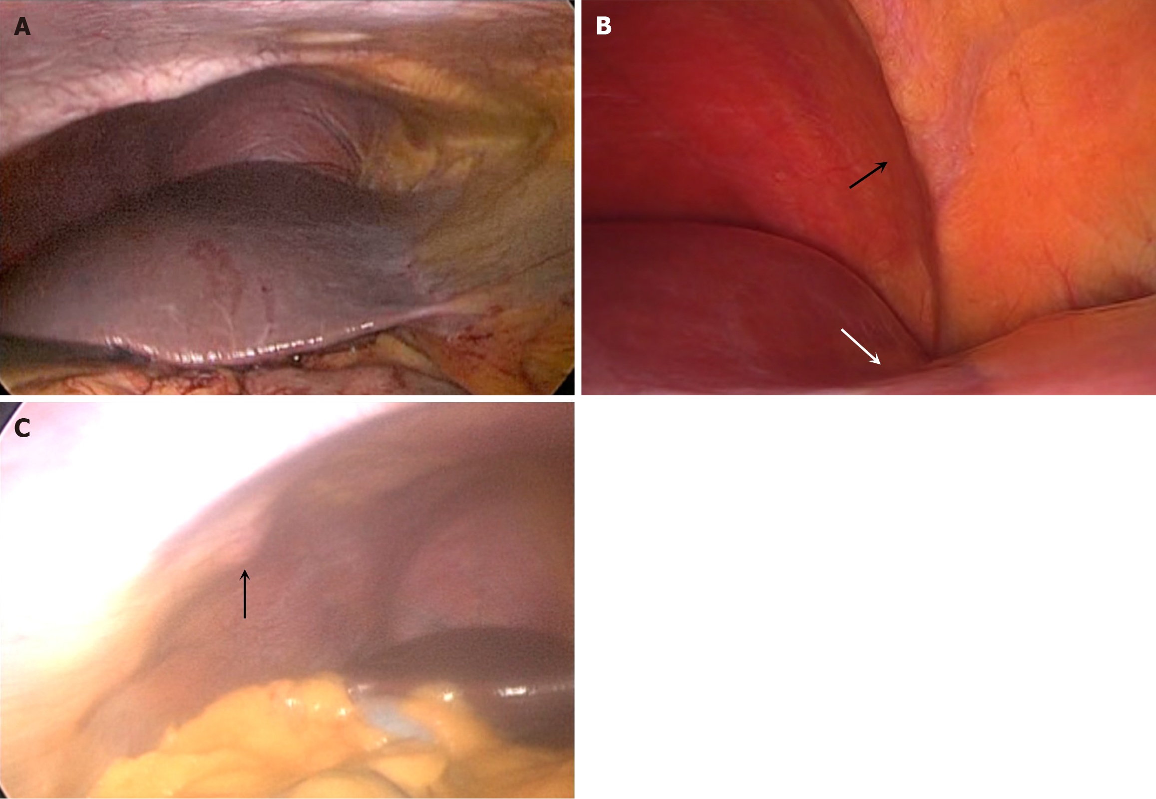
Figure 1 Laparoscopic view.
A: Laparoscopic view of the sub-phrenic space. There is normal anatomy present with a smooth, rounded liver surface contacting a normal diaphragm; B: Laparoscopic view of the right sub-phrenic space. There is a well-defined diaphragmatic band (black arrow) that corresponds to a depression (white arrow) on the surface of segment VIII of the liver; C: Laparoscopic view of the sub-phrenic space, with a rib projection (arrow) present. Note the normal smooth appearance of the liver surface.
- Citation: Cawich SO, Thomas DA, Mohammed F, Gardner MT, Craigie M, Johnson S, Kedambady RS. Hepatic grooves: An observational study at laparoscopic surgery. World J Exp Med 2024; 14(2): 94357
- URL: https://www.wjgnet.com/2220-315x/full/v14/i2/94357.htm
- DOI: https://dx.doi.org/10.5493/wjem.v14.i2.94357


