Copyright
©The Author(s) 2024.
World J Exp Med. Jun 20, 2024; 14(2): 92157
Published online Jun 20, 2024. doi: 10.5493/wjem.v14.i2.92157
Published online Jun 20, 2024. doi: 10.5493/wjem.v14.i2.92157
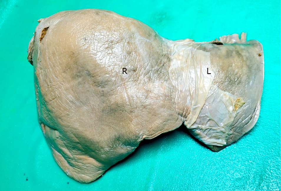
Figure 1 Anatomic specimen of a human liver with conventional surface anatomy.
Both right (R) and left (L) hemi-livers have a smooth, convex surface in contact with the diaphragm.
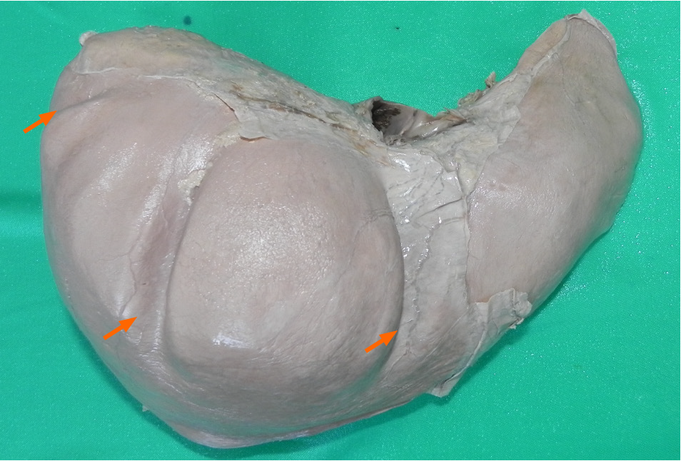
Figure 2 Anatomic specimen of a human liver with variant surface anatomy.
Multiple surface depressions (arrows) are demonstrated on the diaphragmatic surface of liver.
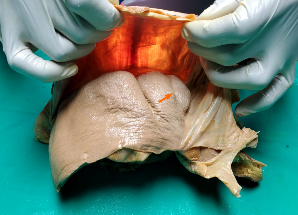
Figure 3 Our technique for observation by trans-illumination.
A light source is applied to the thoracic surface of the diaphragm, allowing hypertrophic muscular bands to be identified (arrow).
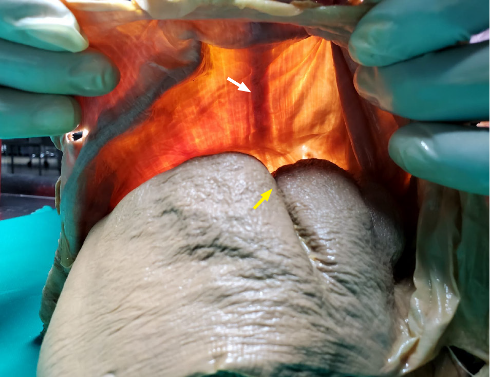
Figure 4 The trans-illumination technique allows hypertrophic muscular bands to be demonstrated (white arrows) in the diaphragm corresponding to surface depressions on the liver (yellow arrows).
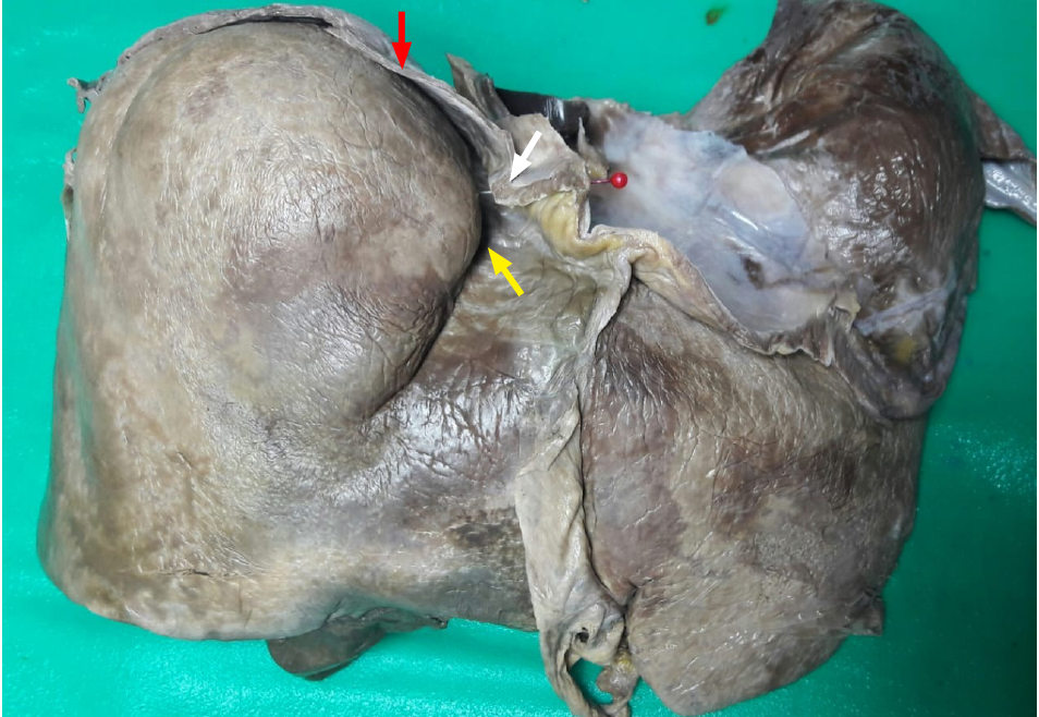
Figure 5 The thickness of the diaphragm is greater (white arrow) adjacent to the surface grooves (yellow arrow), and becomes thin at the smooth liver surface (red arrow).
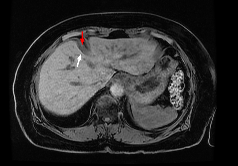
Figure 6 Magnetic resonance imaging of a patient’s abdomen.
There is a deep surface depression in the right hemi-liver (white arrow) and thickened diaphragmatic band (red arrow) can be seen intimately related to the depression.
- Citation: Cawich SO, Gardner MT, Shetty R, Louboutin JP, Dabichan Z, Johnson S. Liver surface depressions in the presence of diaphragmatic muscular bands on trans-illumination. World J Exp Med 2024; 14(2): 92157
- URL: https://www.wjgnet.com/2220-315x/full/v14/i2/92157.htm
- DOI: https://dx.doi.org/10.5493/wjem.v14.i2.92157


