Copyright
©The Author(s) 2019.
World J Exp Med. Dec 19, 2019; 9(2): 14-31
Published online Dec 19, 2019. doi: 10.5493/wjem.v9.i2.14
Published online Dec 19, 2019. doi: 10.5493/wjem.v9.i2.14
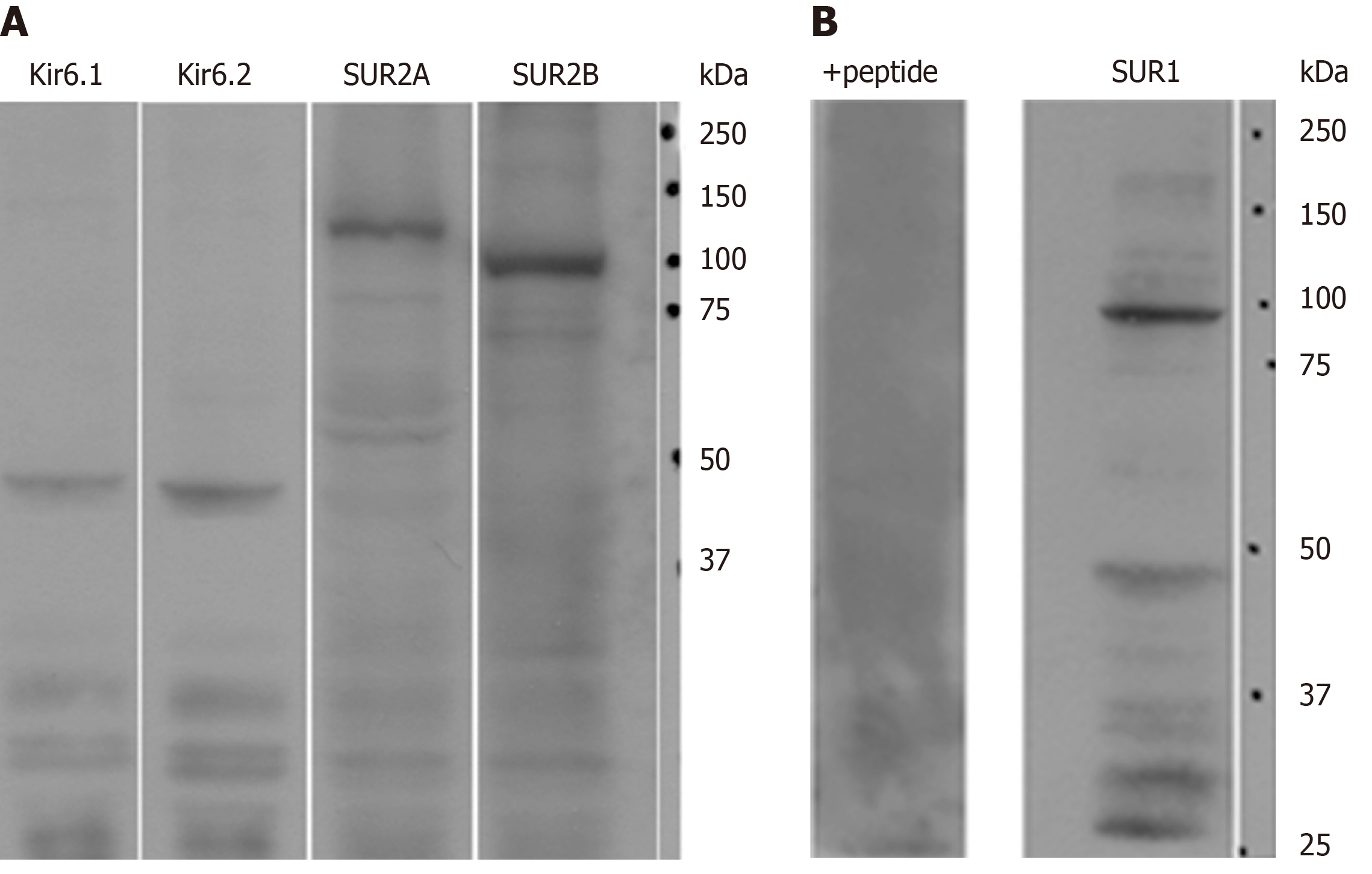
Figure 1 The expression of ATP-sensitive K+ channel subunits in rat liver extracts.
A: Antibodies to Kir6.1, Kir6.2, SUR2A and/or SUR2B showed prominent specific bands, respectively; B: A prominent band around 100 kDa was recognized with our new anti-SUR1 antibody. The band disappeared when blocked by preincubation with the corresponding SUR1 peptide antigen from which the antibody had been raised (+peptide).
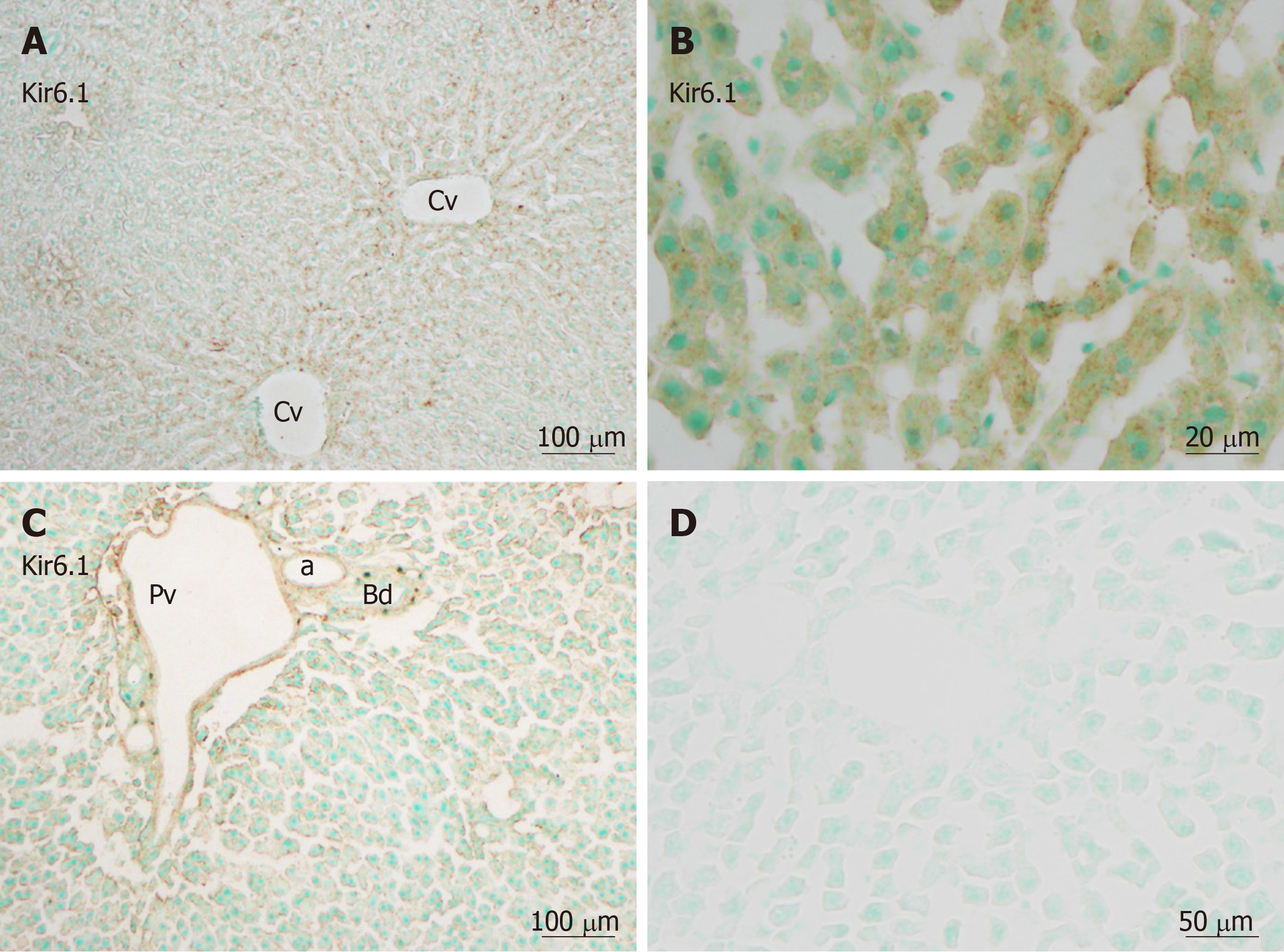
Figure 2 Kir6.
1 immunoreactivity in liver sections. Kir6.1 was expressed in hepatocytes and sinusoidal cells of the liver. A: Staining was intense in the area around the central vein; B: Showing punctate reactivity around the nucleus in hepatocytes cells and sinusoidal cells when viewing with high magnification; C: Weaker immunoreactivity was evident in the triangular areas of the vein and artery, but no reactivity was evident in the bile duct; D: A negative control section devoid of staining. a: Artery; Bd: Bile duct, Cv: Central vein; Pv: Portal vein. Bars: 100 µm (A and C), 20 µm (B), 50 µm (D).
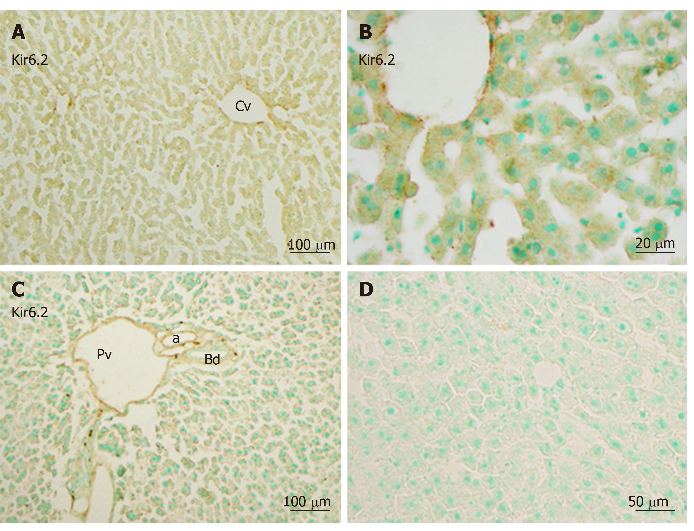
Figure 3 Immunoreactivity with Kir6.
2 was expressed in hepatocytes and sinusoidal cells of the liver. A and B: Mediated immunoreactivity was seen in the area around the central vein; A and C: Mediated immunoreactivity was weaker in the border areas; D: A negative control section devoid of staining (no more than background staining was observed). a: Artery; Bd: Bile duct, Cv: Central vein; Pv: Portal vein. Bars: 100 µm (A and C), 20 µm (B), 50 µm (D).
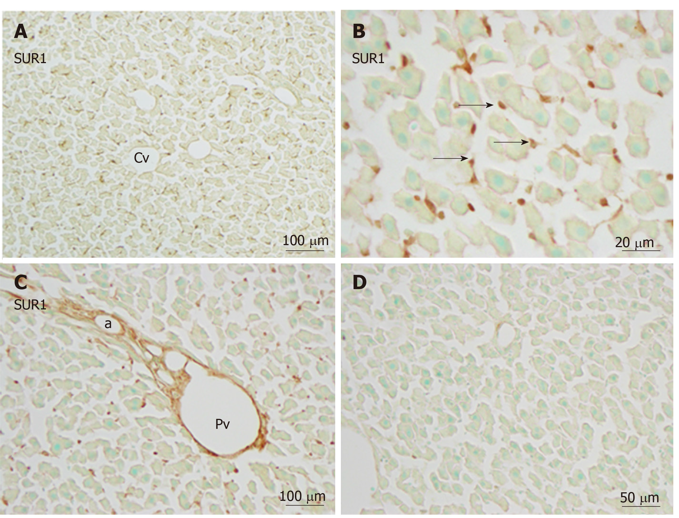
Figure 4 Immunoreactivity with SUR1 was mainly evident in sinusoidal cells (arrows) and weaker in the cell membranes of hepatocytes.
A and B: There was no such tendency toward immunoreactivity with SUR1 in the area of the central vein vs the border area; C: In the portal area, endothelial cells of the blood vessels showed intense immunoreactivity with SUR1; D: Absorption negative control section devoid of staining (no more than background) after incubation with cognate antigen peptide. a: Artery, Cv: Central vein; Pv: Portal vein. Bars: 100 µm (A and C), 20 µm (B), 50 µm (D).
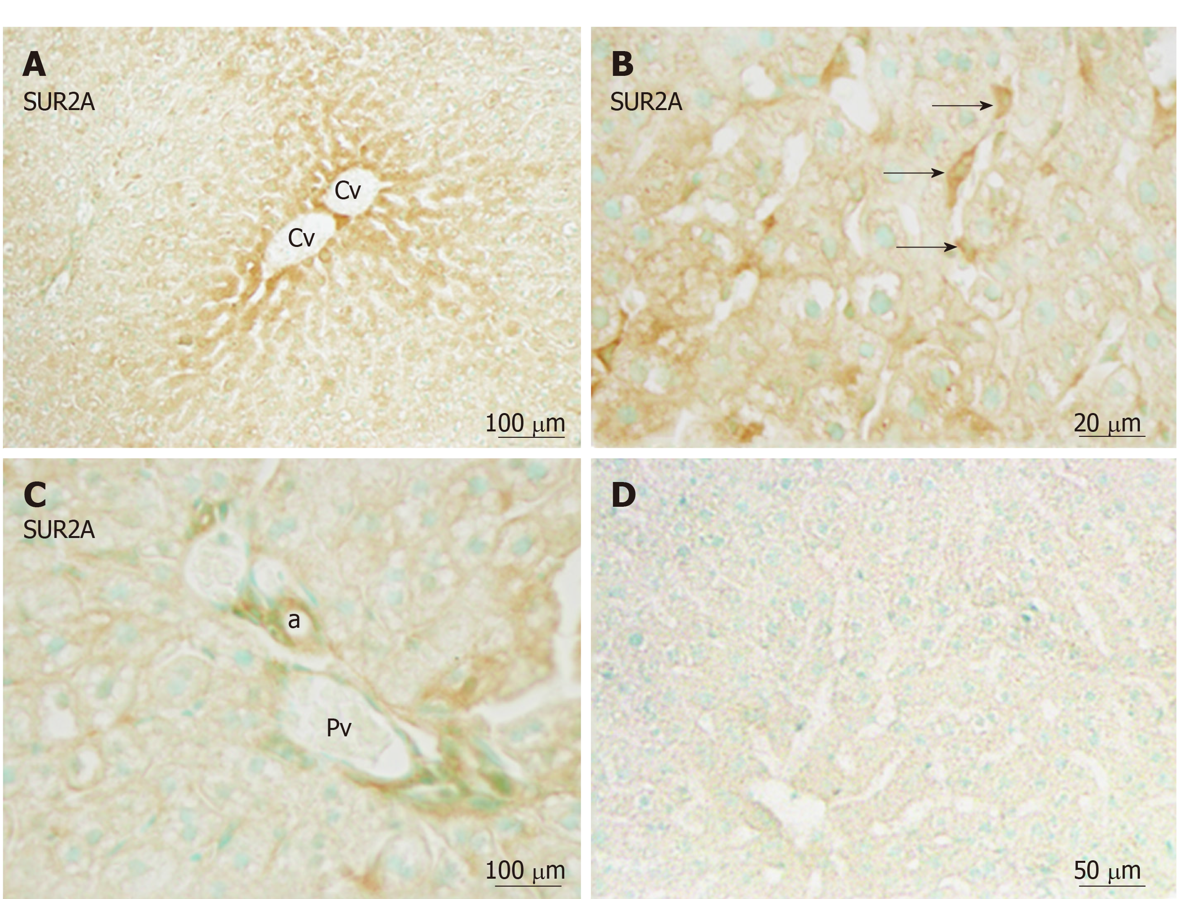
Figure 5 Immunoreactivity with SUR2A was evident in hepatocytes and sinusoidal cells (arrows) of the liver.
A: Immunoreactivity with SUR2A was stronger in the area of the central vein (Cv) vs the border areas; B: Weaker immunoreactivity in the cell membrane and moderate immunoreactivity in the sinusoidal cells were observed (arrows); C: Weaker immunoreactivity with SUR2A was also observed in the endothelial cells of the blood vessels; D: Absorption negative control section devoid of staining (no more than background was observed). a: Artery. Cv: Central vein; Pv: Portal vein. Bars: 100 µm (A and C), 20 µm (B), 50 µm (D).
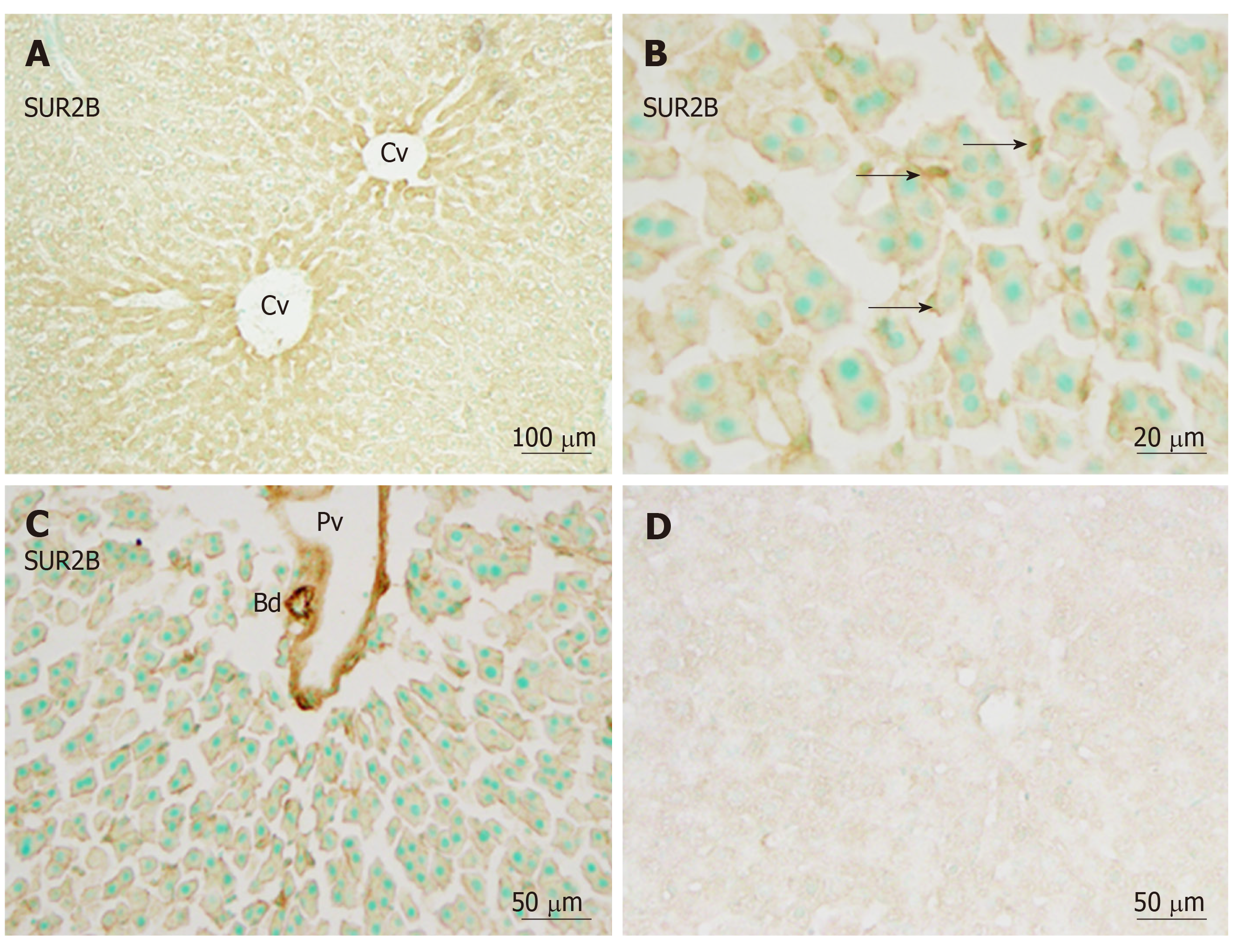
Figure 6 Immunoreactivity with SUR2B.
A: Immunoreactivity with SUR2B was stronger in the area of the central vein (Cv) vs the border areas; B: Immunoreactivity with SUR2B was expressed in hepatocytes and sinusoidal cells (arrows) of the liver. Weaker immunoreactivity in the cell membrane and medium immunoreactivity in sinusoidal cells was observed (arrows); C: Immunoreactivity with SUR2B was also observed in the endothelial cells of blood vessels and bile ducts; D: Absorption negative control section devoid of staining (no more than background was observed). Bd: Bile duct; Cv: Central vein; Pv: Portal vein. Bars: 100 µm (A), 20 µm (B), 50 µm (C and D).
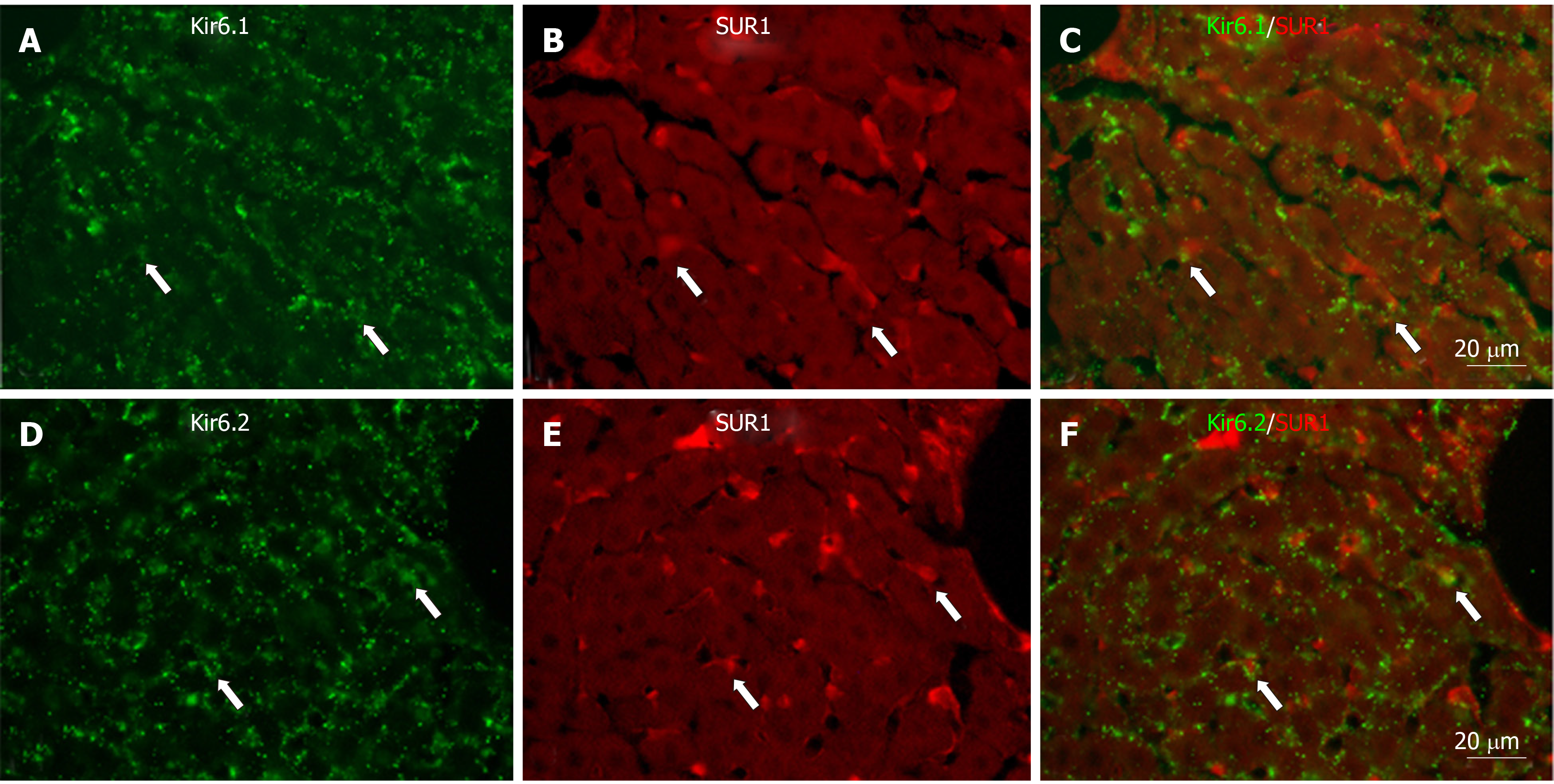
Figure 7 Immunofluorescence double staining for Kir6.
1 and/or Kir6.2 with SUR1 in liver sections. A and D: Kir6.1 and Kir6.2 were widely localized in hepatocyte and sinusoidal cells; B and E: SUR1 was mainly localized in sinusoidal cells; C: There were few sinusoidal cells that had colocalized Kir6.1 and SUR1 (arrows); F: There were few sinusoidal cells that had colocalized Kir6.2 and SUR1 (arrows). Bars: 20 µm.
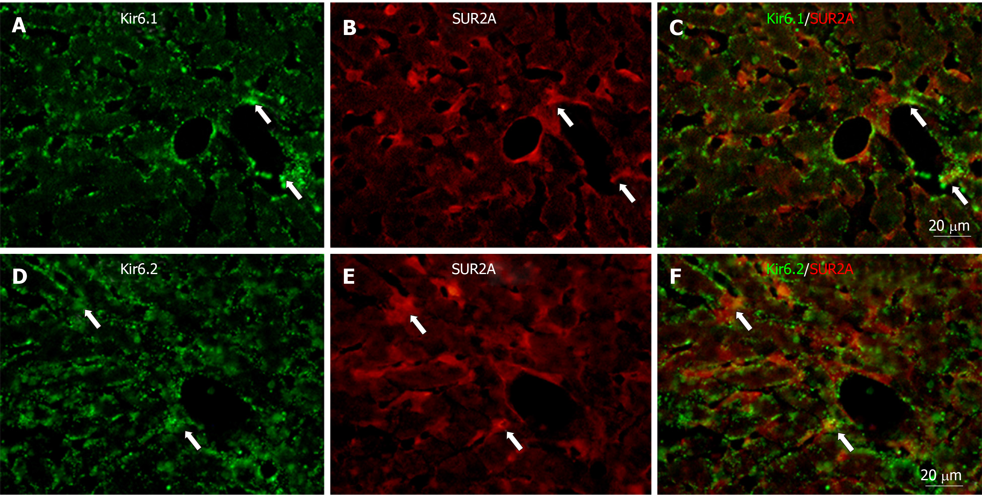
Figure 8 Immunofluorescence double staining for Kir6.
1 and/or Kir6.2 with SUR2A in liver sections. A and D: Kir6.1 and Kir6.2 were widely localized in hepatocyte and sinusoidal cells; B and E: SUR2A was mainly localized in sinusoidal cells; C: There were few sinusoid cells with colocalized Kir6.1 and SUR2A (arrows); F: There were few sinusoidal cells with colocalized Kir6.2 and SUR2A (arrows). Bars: 20 µm.
Figure 9 Immunofluorescence double staining for Kir6.
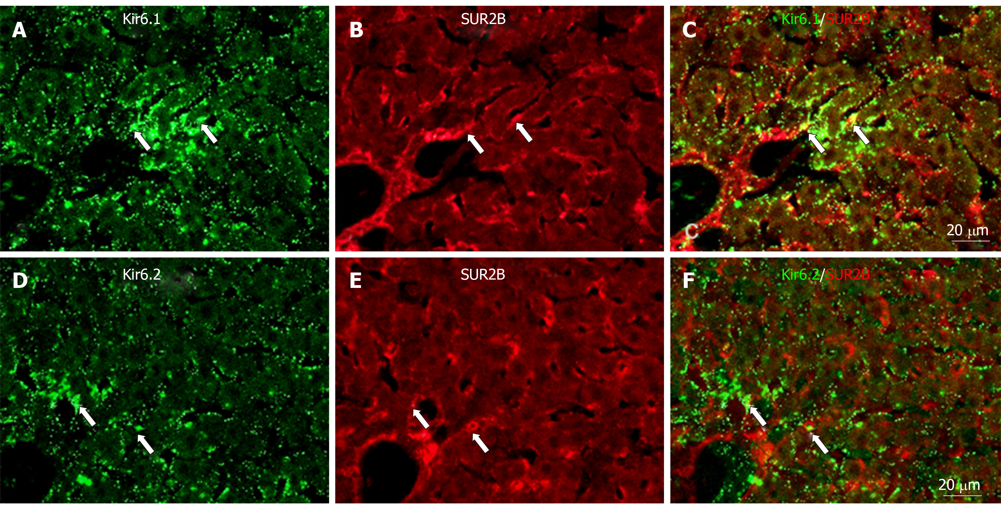
1 and/or Kir6.2 with SUR2B in liver sections. A and D: Kir6.1 and Kir6.2 were widely localized in hepatocyte and sinusoidal cells; B and E: SUR2B staining was mainly present in sinusoidal cells; C: There were few sinusoidal cells with colocalized Kir6.1 and SUR2B (arrows); F: There were few sinusoid cells with colocalized Kir6.2 and SUR2B (arrows). Bars: 20 µm.
Figure 10 Immunofluorescence double staining.
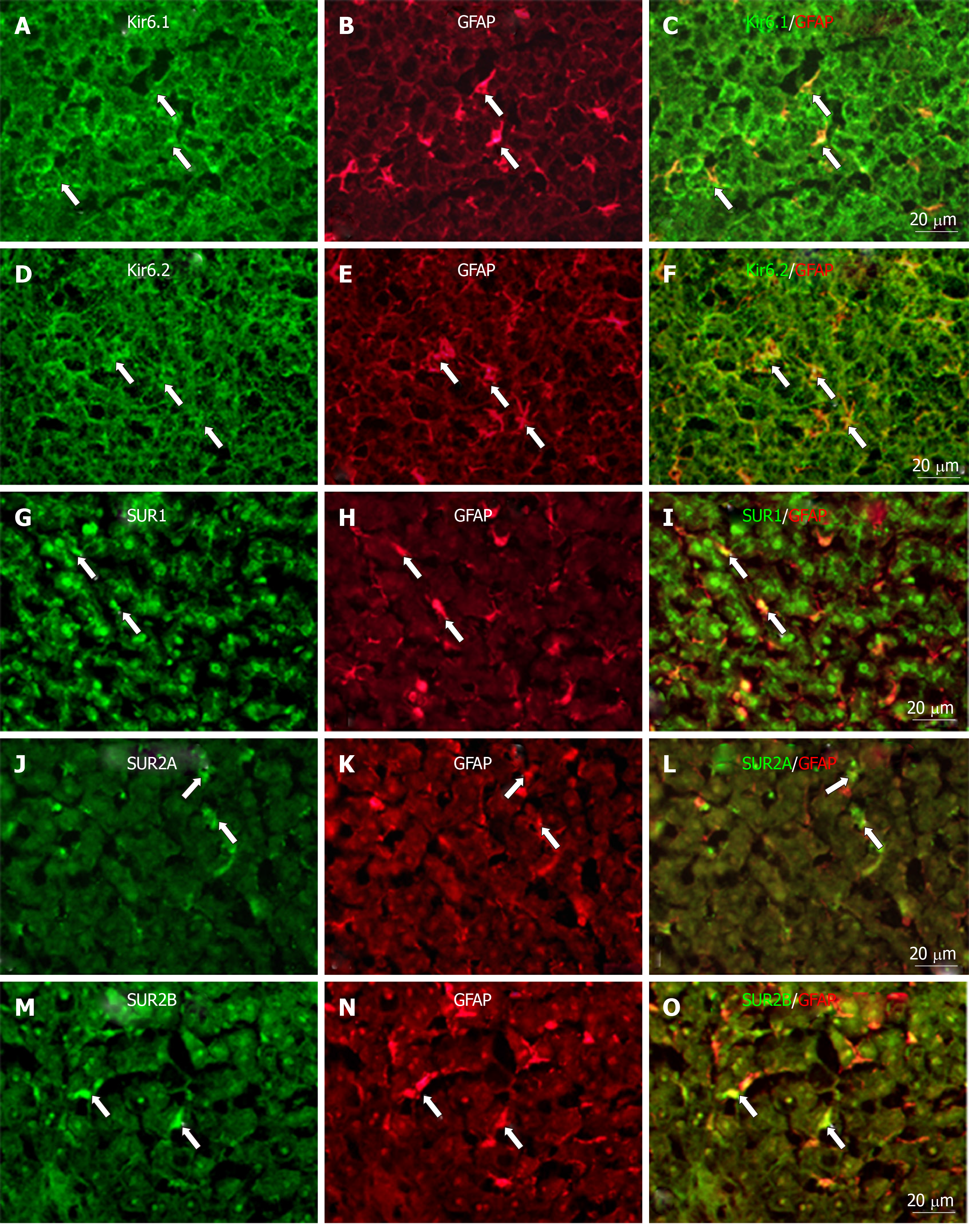
Immunofluorescence double staining for Kir6.1, Kir6.2, SUR1, SUR2A, and/or SUR2B (A, D, G, J, and M) and GFAP [B, E, H, K, and N, marker of hepatic stellate cells (HSCs)] in no-fixed liver sections; Most of the GFAP+ cells (HSCs) colocalized with Kir6.1 (C, arrows) and/or Kir6.2 (F, arrows); There were few GFAP+ cells (HSCs) colocalized with SUR1 (i, arrows), SUR2A (L, arrows), and/or SUR2B (O, arrows). Bars: 20 µm.
Figure 11 Immunofluorescence double staining for ATP-sensitive K+ channel subunits Kir6.
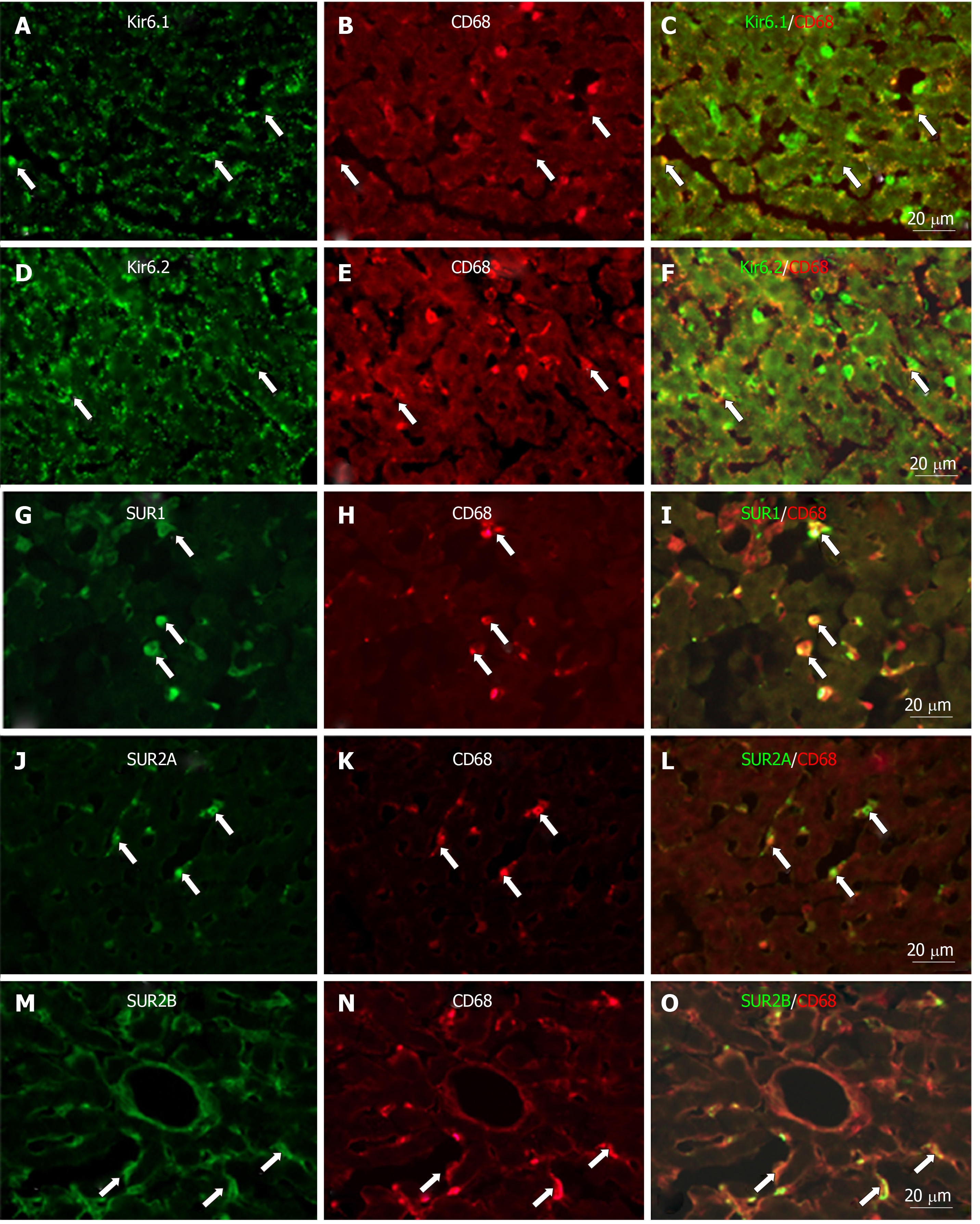
1, Kir6.2, SUR1, SUR2A and SUR2B with CD68 in liver sections. Immunofluorescence double staining for ATP-sensitive K+ channel subunits Kir6.1 (A), Kir6.2 (D), SUR1 (G), SUR2A (J) and SUR2B (M) with CD68 (B, E, H, K, and N, marker of Kupffer cells) in liver sections; Some CD68+ cells (Kupffer cells) colocalized with Kir6.1 (C, arrows) and/or Kir6.2 (F, arrows), but most colocalized with SUR1 (I, arrows), SUR2A (L, arrows) and SUR2B (O, arrows). Bars: 20 µm.
Figure 12 Immunofluorescence double staining for ATP-sensitive K+ channel subunits Kir6.
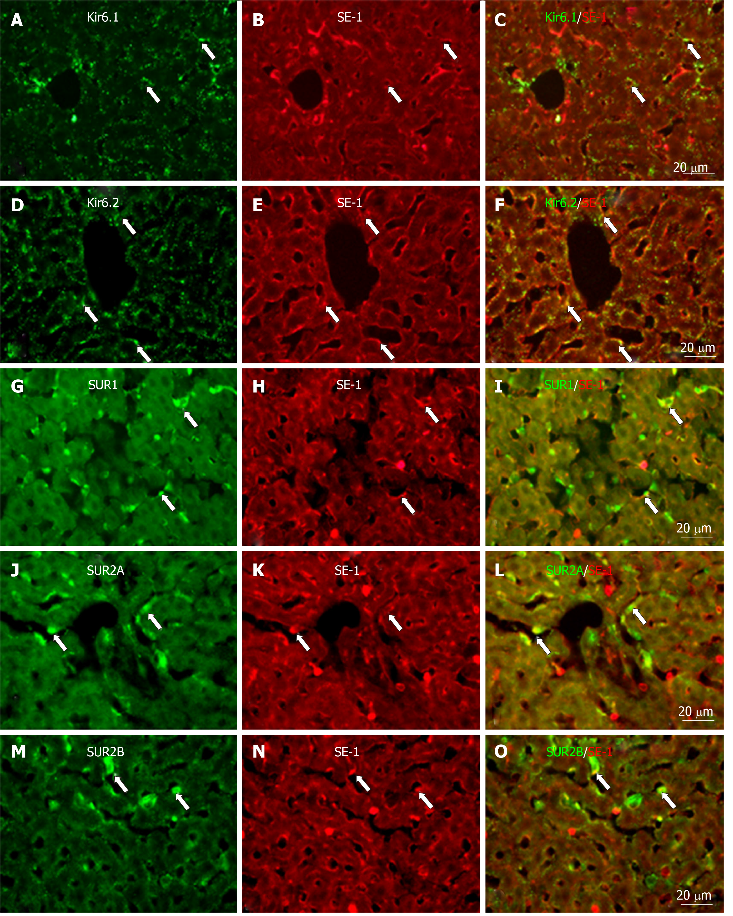
1, Kir6.2, SUR1, SUR2A and SUR2B with SE-1. Immunofluorescence double staining for ATP-sensitive K+ channel subunits Kir6.1, Kir6.2, SUR1, SUR2A and SUR2B (A, D, G, J, and M) with SE-1 [marker of sinusoidal endothelial cells (SECs)]; In the liver sections, SECs showed immunopositive reactivity to SE-1 (B, E, H, K, and N); A few SE-1+ cells colocalized with Kir6.1 (C, arrows), but a number of them colocalized with Kir6.2 (F, arrows); As for SURs, some SE-1+ cells colocalized with SUR1 (I, arrows) and/or SUR2B (O, arrows); Most SE-1+ cells colocalized with SUR2A (L, arrows). Bars: 20 µm.
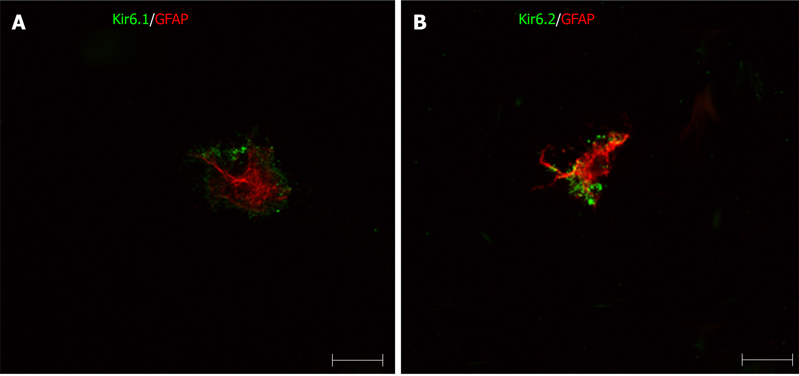
Figure 13 Immunofluorescence double staining for pore-forming subunits in primary cultured hepatic stellate cells.
The primary hepatic stellate cells were marked by GFAP (red), they showed immunoreactivity to Kir6.1 and Kir6.2 (green). Bars: 50 µm.
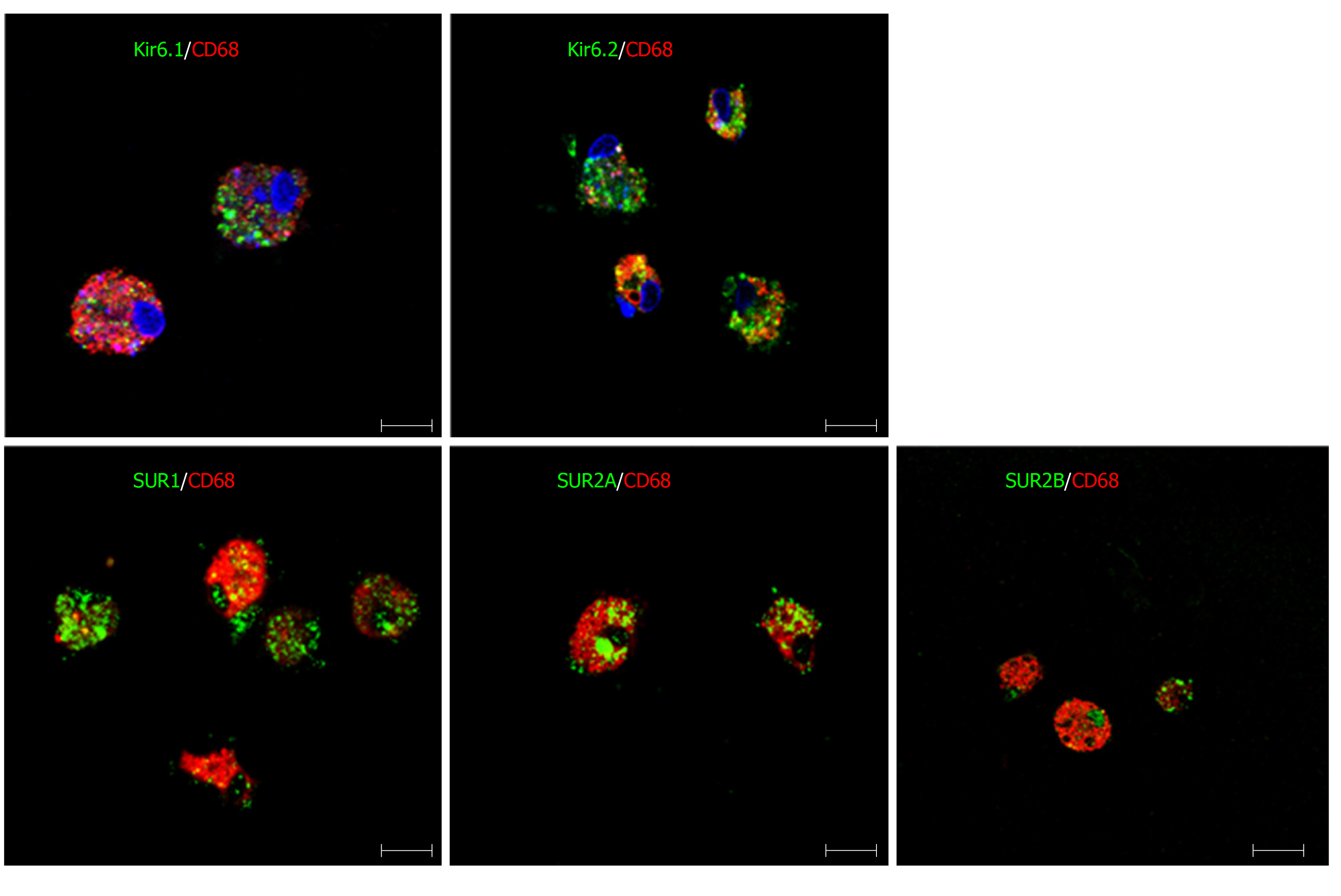
Figure 14 Immunofluorescence double staining for ATP-sensitive K+ channel subunits in primary cultured Kupffer cells.
Primary Kupffer cells were labeled with CD68 (red). These CD68+ Kupffer cells also showed immunoreactivity to five kinds of ATP-sensitive K+ channel subunits: Kir6.1, Kir6.2, SUR1, SUR2A and SUR2B (green). Bars: 50 µm.
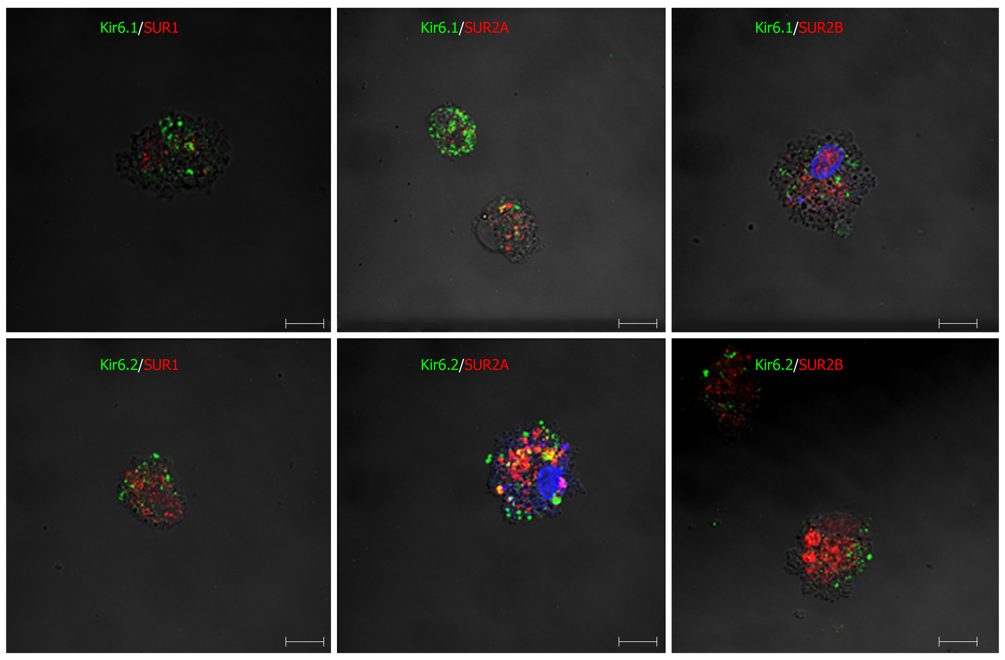
Figure 15 Immunofluorescence double staining for combinations of pore-forming subunits and regulatory subunits in primary cultured Kupffer cells.
In primary Kupffer cells, the pore-forming subunits Kir6.1 and Kir6.2 are labeled in green, and the regulatory subunits SUR1, SUR2A and SUR2B are labeled in red. The combinations of Kir6.1/SUR1, Kir6.1/SUR2A, Kir6.1/SUR2B, Kir6.2/SUR1, Kir6.2/SUR2A and Kir6.2/SUR2B are all exhibited in the Kupffer cells. Bars: 50 µm.
- Citation: Zhou M, Yoshikawa K, Akashi H, Miura M, Suzuki R, Li TS, Abe H, Bando Y. Localization of ATP-sensitive K+ channel subunits in rat liver. World J Exp Med 2019; 9(2): 14-31
- URL: https://www.wjgnet.com/2220-315X/full/v9/i2/14.htm
- DOI: https://dx.doi.org/10.5493/wjem.v9.i2.14


