Copyright
©The Author(s) 2016.
World J Exp Med. Nov 20, 2016; 6(4): 72-79
Published online Nov 20, 2016. doi: 10.5493/wjem.v6.i4.72
Published online Nov 20, 2016. doi: 10.5493/wjem.v6.i4.72
Figure 1 Differentiation of neuronal progenitor cells into neuronal cells, oligodendrocytes and astrocytes under in vitro culture conditions.
A: Neuron; B: Oligodendrocytes; C: Astrocytes.
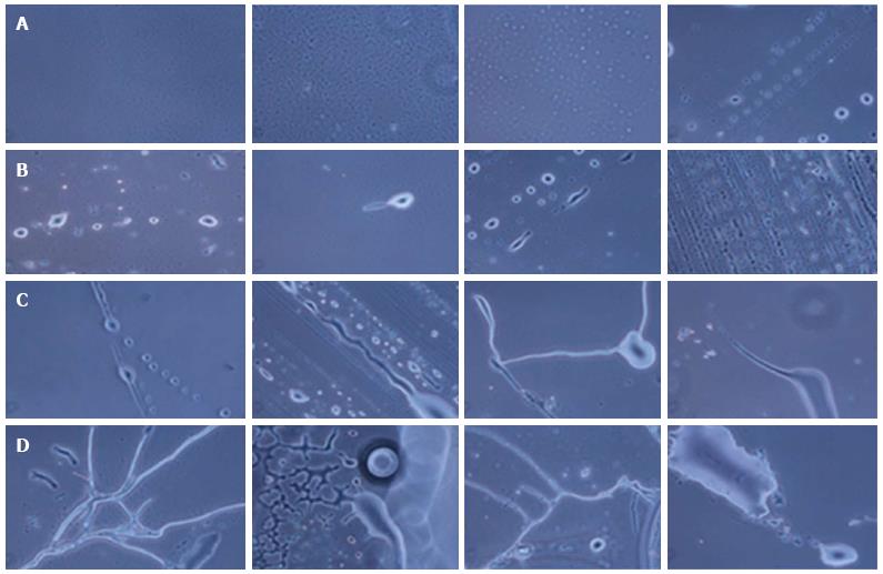
Figure 2 Phase contrast images of neuronal cells and tissues at different days of culture.
A: Day 0: Small stem cells; B: Day 5: Differentiating neurons; C: Day 16-18: Small protrusions, neuritis structure and end-joining of neurons; D: Day 30: Complete neurons and neuronal tissue.
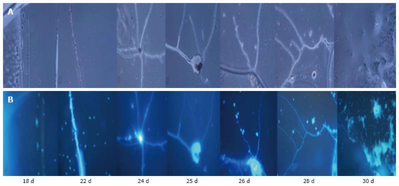
Figure 3 4’, 6-diamidino-2-phenylindole staining of neuronal specific cells and tissues under in vitro culture condition.
A: Cells viewed under a phase contrast microscope; B: 4’6-diamidino-2-phenylindole stained cells.
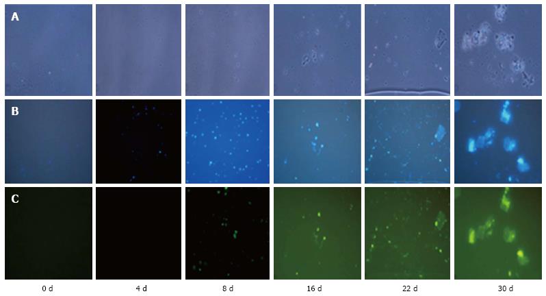
Figure 4 Differentiation of human embryonic stem cells into neuronal specific cells and tissues under in vitro culture conditions.
A: Cells viewed under a phase contrast microscope; B: 4’, 6-diamidino-2-phenylindole stained cells; C: Green (NeuN) stained cells.
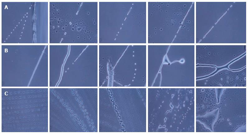
Figure 5 Mechanism of differentiation of human embryonic stem cells into neurons and axons formation under in vitro culture conditions.
A: Day 5: Neural progenitor cells increased in size, arranged in a line and then joined together through small protrusions; B: Day 16-18: Cytoplasmic material mixed together to form long axonal structure; C: Day 30: Formation of neuronal tissue.
- Citation: Shroff G. Morphogenesis of human embryonic stem cells into mature neurons under in vitro culture conditions. World J Exp Med 2016; 6(4): 72-79
- URL: https://www.wjgnet.com/2220-315X/full/v6/i4/72.htm
- DOI: https://dx.doi.org/10.5493/wjem.v6.i4.72


