Copyright
©The Author(s) 2016.
World J Exp Med. Nov 20, 2016; 6(4): 63-71
Published online Nov 20, 2016. doi: 10.5493/wjem.v6.i4.63
Published online Nov 20, 2016. doi: 10.5493/wjem.v6.i4.63
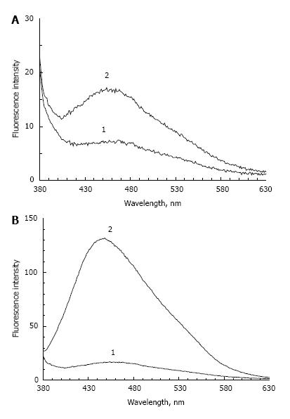
Figure 1 Fluorescent products accumulation in weakly oxidised bovine photoreceptor outer segments after 72 h incubation at 37 °C.
A: POS fluorescence incubated in the absence of fructose: curve 1 - prior to incubation; curve 2 - after incubation; B: POS fluorescence incubated in the presence of fructose: curve 1 - prior to incubation; curve 2 - after incubation. Fluorescence was measured in 0.1 mol/L K-phosphate buffer, pH 7.6, excitation wavelength - 365 nm.
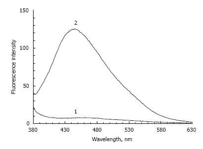
Figure 2 Fluorescent products accumulation in the pre-oxidized photoreceptor outer segments at 72 h incubation at 37 °C.
Curve 1 - prior to incubation; curve 2 - after incubation. Measurement conditions as in the legend under Figure 1.
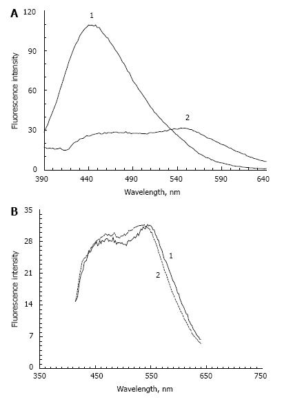
Figure 3 Production of model lipofuscin in the reaction between modified photoreceptor outer segments and bisretinoid A2E.
A: A2E binding by modified photoreceptor outer segments leads to a change in their fluorescence characteristics: curve 1 - POS modified oxidized prior to incubation with A2E; curve 2 - modified POS containing A2E (LPO - lipofuscin); B: Comparison of fluorescence characteristics of the model lipofuscin (LPO) and natural lipofuscin granules from human RPE: curve 1 (solid line) - LPO; curve 2 (dotted line) - lipofuscin granules from human RPE. Model lipofuscin preparation is described in materials and methods, excitation wavelength - 365 nm. POS: Photoreceptor outer segments; RPE: Retinal pigment epithelium.
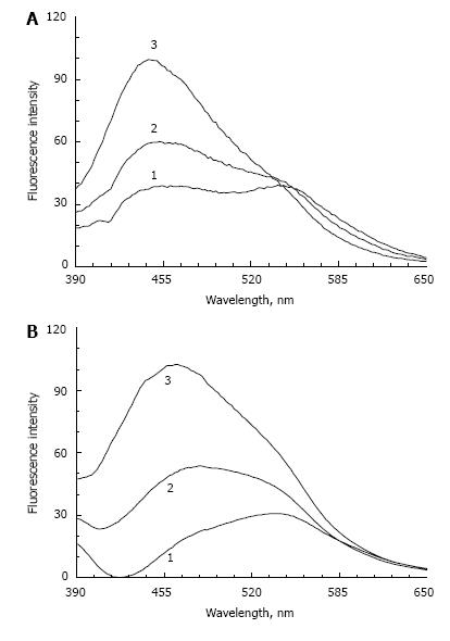
Figure 4 Effect of visible light irradiation on the fluorescence characteristics of lipofuscin.
A: Change of fluorescence spectra of the model lipofuscin (LFF): 1 - prior to irradiation, 2 and 3 - after irradiation for 1 and 3 h, respectively; B: Change of fluorescence spectra of human RPE lipofuscin granules: 1 - prior to irradiation, 2 and 3 - after irradiation for 1 and 3 h, respectively. RPE: Retinal pigment epithelium.
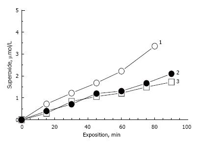
Figure 5 Comparative kinetics of superoxide generation by irradiation of natural lipofuscin (curve 1) and model of lipofuscin (curve 2 - LFF, curve 3 - LPO).
The reaction conditions - see methods. Lipofuscin concentrations - 2.5 mg/mL.
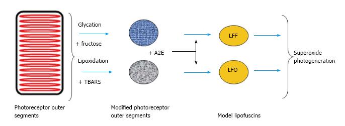
Figure 6 Schema of processes generation of retinal pigment epithelium lipofuscin from modified photoreceptor outer segments and fluorophore A2E.
POS: Photoreceptor outer segments; TBARS: TBA-reactive substances.
- Citation: Dontsov A, Koromyslova A, Ostrovsky M, Sakina N. Lipofuscins prepared by modification of photoreceptor cells via glycation or lipid peroxidation show the similar phototoxicity. World J Exp Med 2016; 6(4): 63-71
- URL: https://www.wjgnet.com/2220-315X/full/v6/i4/63.htm
- DOI: https://dx.doi.org/10.5493/wjem.v6.i4.63


